
Page 318 - IJB-9-2
P. 318
International Journal of Bioprinting Flexible 3D printing in cardiovascular medicine
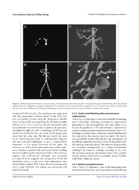
Figure 2. Results of segmentation and 3D post-processing. (A) Segmentation after removing all non-vascular structures and smoothing. (B) Surface model
generated by stack triangulation using the integrated 3D Viewer plugin. (C) Vascular model after using the “Bisect” tool to remove unnecessary arteries and
cut the vessels open in Blender. (D) Vascular model after generating vascular walls with 1-mm wall thickness (white arrow heads).
Kommerell’s diverticulum. The aneurysm was segmented 3.2.2. Active renal bleeding after percutaneous
with the open-source software based on the CTA data nephrostomy
and successfully printed using the transparent, flexible Case 2 (Figure 4) illustrates a case of new onset life-threatening
resin. As the model was larger than the 3D printer’s build renal hemorrhage following percutaneous nephrostomy
volume (14.5 × 14.5 × 18.5 cm), Blender was used to slice placement to treat hydronephrosis and renal failure in an
the model into two parts in order to print them separately. 87-year-old gentleman. CTA demonstrated a nephrostomy
An ultraviolet light pen with a wavelength of 405 nm was catheter running along the segmental renal artery (Figure 3A)
used to fuse both parts into one aortic model using liquid resulting in vascular injury and active arterial bleeding into
resin from the resin tank. The life-size model was used the renal pelvis, thus causing macrohematuria. The patient
by an interdisciplinary team of cardiovascular surgeons was successfully treated with selective coil embolization of
and interventional radiologists to gain a preprocedural the affected segmental renal artery at our IR department, and
impression of the actual dimension of the aorta, the the bleeding eventually stopped. The endovascular procedure
aneurysm, as well as the location and course of the supra- was simulated retrospectively by a young interventional
aortic arteries in regard to the aneurysm, thereby assisting radiologist for hands-on training according to the real-
the team in planning the complex procedure. The combined life procedure using the 3D-printed model of the patient’s
surgical and endovascular treatment was successful visceral arteries connected to a peristaltic pump (Guerbet
in a total of three surgeries: the transposition of the left KMP 2000, Villepinte, France).
subclavian artery, a total aortic arch replacement with
frozen elephant trunk (FET), and a thoracic endovascular 3.2.3. Splenic pseudoaneurysm
aortic repair (TEVAR). The patient was discharged well Case 3 (Figure 5) illustrates a case of life-threatening acute
from the hospital. arterial hemorrhage from splenic pseudoaneurysm following
Volume 9 Issue 2 (2023) 310 https://doi.org/10.18063/ijb.v9i2.669