
Page 317 - IJB-9-2
P. 317
International Journal of Bioprinting Flexible 3D printing in cardiovascular medicine
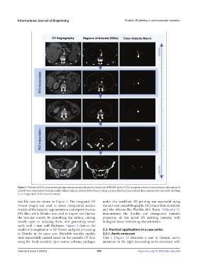
Figure 1. Process of ROI interpolation in segmenting vascular structures. Based on a DICOM stack of CTA images in arterial contrast phase, the regions of
interest were interpolated from manually defined regions (yellow dotted lines). Using a macro, the structures outside these regions were removed, resulting
in an image stack of the visceral arteries.
real-life case are shown in Figure 1. The integrated 3D under this workflow. 3D printing was successful using
Viewer plugin was used to create triangulated surface the end-user stereolithography 3D printer from Formlabs
models of the vascular segmentations and export them as and the silicone-like Flexible 80A Resin. Videoclip S1
STL files, while Blender was used to import and finalize demonstrates the flexible and transparent material
the vascular models by smoothing the surface, cutting properties of this novel 3D printing material with
vessels open or reducing them, and generating vessel biological tissue mimicking characteristics.
walls with 1-mm wall thickness. Figure 2 depicts the
results of triangulation in 3D Viewer and post-processing 3.2. Practical applications in a case series
in Blender in the same case. Printable vascular models 3.2.1. Aortic aneurysm
were successfully created based on the patient’s CT data Case 1 (Figure 3) illustrates a case of thoracic aortic
using the freely available open-source software packages aneurysm in the right descending aorta associated with
Volume 9 Issue 2 (2023) 309 https://doi.org/10.18063/ijb.v9i2.669