
Page 408 - IJB-9-2
P. 408
International Journal of Bioprinting 3D-printed skin substitute accelerates wound healing in vivo
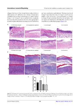
collagen filled most of the dermal defects without obvious not been completely re-epithelialized. The skin structure of
inflammatory cell infiltration, but obvious inflammatory Group B was better than that of Groups C and D and was
cell infiltration was observed in Groups A, C, and D. Neither similar to that of Group A. The proliferation of collagen
Group C nor Group D was re-epithelialized completely. in Group B had completely filled the dermal defect, and
Fourteen days after operation, the other three groups have the degree of inflammatory cell infiltration was less severe
formed complete skin structures except Group D which has than that in the other three groups (Figure 5A).
A
B
C
Figure 5. (A) Hematoxylin-eosin staining was performed to evaluate the effect of different methods on histology of wound healing on days 7 and 14 after
the operation. Scale bar = 100 μm. (B) Masson staining was performed to observe the effect of different methods on the production and arrangement of
collagen on day 14. Scale bar = 100 μm. (C) The area percentage of collagen in each group was quantified. **P < 0.01.
Volume 9 Issue 2 (2023) 400 https://doi.org/10.18063/ijb.v9i2.674