
Page 42 - IJB-9-2
P. 42
International Journal of Bioprinting Scaffolds printed with light sheet stereolithography
A B
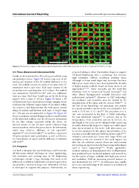
Figure 4. Fluorescence images of microsphere pipetted inside the scaffold with a pore size of (A) 68 ± 2.5 μm and (B) 149.9 ± 2.3 μm.
3.3. Three-dimensional interconnectivity propose looking at other illumination shapes to support
Finally, we demonstrated the 3D printing of scaffolds using VP-based bioprinting with a technology that provides
our prototype device. Figure 5A shows a top view of the high resolution without sacrificing printing areas.
microscopic structure of the 3D scaffold fabricated in this Although its linear voxel shape may constrain free-form
fabrication, many studies have demonstrated the benefits
work. The scaffold consists of a series of six layers with 45° of highly porous scaffolds based on linear struts to cell
orientation within each other. Each layer consists of 100 regeneration [9,13,45] . Some examples are the mesh-like
struts that took a printing time of 3.5 s/layer. The scaffold structures used in commercial wound dressings and
[13]
has orientations 0/45/90/135/180° with one additional other fibrous bioengineered scaffolds fabricated with
layer as a base. Each layer height was set by the building sophisticated methods . However, in DLP technology,
[25]
support with a value of 100 μm. Figure 5B shows a 3D the resolution and projection area are limited by the
reconstruction from confocal microscopy imaging, which magnification of the optics and the current DMD . To
[56]
evidences the different height values of the layers within the best of our knowledge, the maximum area printed
the construct and demonstrates the void spaces created at a lateral resolution similar to the size achieved in this
along the surface and between each layer. A height range work is 19.35 mm × 12.1 mm . The projection area in
[41]
of ≈ 526 μm was reconstructed in Figure 5B. Figure 5C that case corresponds to the limit of DMD technology
shows a computer-assisted design model of a small section for near ultraviolet sources . In contrast, due to the
[57]
of the fabricated scaffold, and the 3D pore is represented independence from projection systems in LS-SLA, the
by the blue volume contained within the struts. An line length of the struts can be enlarged while conserving
internal section of the 3D pore model evidences high its width. Beyond the large strut length and width aspect
interconnectivity within the pores due to 3D fabrication, ratio demonstrated in this work, both dimensions can
which may influence efficiency of cell ingrowth , be further optimized by the optical characteristics of the
[5]
migration , and directionality . In addition, engineered scan lens as initially estimated with Equations 1 and 2 [58,59] .
[25]
[45]
3D pores promote water penetration, as well as influence Available commercial scan lenses with larger FOVs and
the transmission of vapors and the diffusion of nutrients resolution as the one selected in this work support the
and waste . idea that the validated concept allows for improvement
[8]
and scaling-up opportunities for bioprinting technologies
3.4. Prospects
used in tissue engineering [60,61] . Finally, application-
It is hard to imagine that one technology could overcome oriented solutions as demonstrated with LS illumination
all fabrication-related challenges in tissue engineering. may positively boost the health industry and research on
The resolution and printing area in 3D bioprinting the fabrication of larger scaffolds with high pore control
technologies remain a huge challenge that needs to be and resolution. With an increasing research interest on
addressed. In addition, hybrid and customized approaches new biomaterials for VP [32,62] , its techniques may rapidly
can provide a solution to large-scale fabrication. LS-SLA join in as a key technology in the research and industry of
is developed to support this purpose. Alternatively, we tissue engineering.
Volume 9 Issue 2 (2023) 34 https://doi.org/10.18063/ijb.v9i2.650