
Page 80 - IJB-9-2
P. 80
Clinical applications of bioprinted active bone
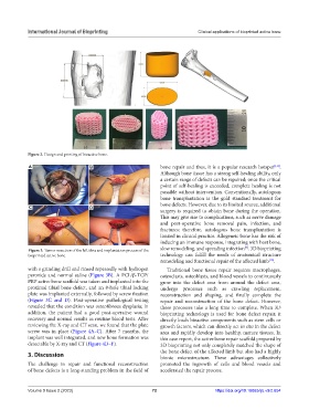
Figure 2. Design and printing of bioactive bone.
A B bone repair and thus, it is a popular research hotspot [1,8] .
Although bone tissue has a strong self-healing ability, only
a certain range of defects can be repaired; once the critical
point of self-healing is exceeded, complete healing is not
possible without intervention. Conventionally, autologous
bone transplantation is the gold standard treatment for
bone defects. However, due to its limited source, additional
C D
surgery is required to obtain bone during the operation.
This may give rise to complications, such as nerve damage
and post-operative bone removal pain, infection, and
fractures; therefore, autologous bone transplantation is
limited in clinical practice. Allogeneic bone has the risk of
inducing an immune response, integrating with host bone,
[9]
Figure 3. Tumor resection of the left tibia and implantation process of the slow remodeling, and spreading infection . 3D bioprinting
bioprinted active bone. technology can fulfill the needs of anatomical structure
remodeling and functional repair of the affected limb .
[10]
with a grinding drill and rinsed repeatedly with hydrogen Traditional bone tissue repair requires macrophages,
peroxide and normal saline (Figure 3B). A PCL/β-TCP/ osteoclasts, osteoblasts, and blood vessels to continuously
PRP active bone scaffold was taken and implanted into the grow into the defect area from around the defect area,
proximal tibial bone defect, and an 8-hole tibial locking undergo processes such as crawling replacement,
plate was implanted externally, followed by screw fixation reconstruction and shaping, and finally complete the
(Figure 3C and D). Post-operative pathological testing repair and reconstruction of the bone defect. However,
revealed that the condition was osteofibrous dysplasia; in these processes take a long time to complete. When 3D
addition, the patient had a good post-operative wound bioprinting technology is used for bone defect repair, it
recovery and normal results in routine blood tests. After directly loads bioactive components such as stem cells or
reviewing the X-ray and CT scan, we found that the plate growth factors, which can directly act in situ in the defect
screw was in place (Figure 4A–C). After 7 months, the area and rapidly develop into healthy, mature tissues. In
implant was well integrated, and new bone formation was this case report, the active bone repair scaffold prepared by
detectable by X-ray and CT (Figure 4D–F). 3D bioprinting not only completely matched the shape of
3. Discussion the bone defect of the affected limb but also had a highly
bionic microstructure. These advantages collectively
The challenge in repair and functional reconstruction promoted the ingrowth of cells and blood vessels and
of bone defects is a long-standing problem in the field of accelerated the repair process.
Volume 9 Issue 2 (2023) 72 https://doi.org/10.18063/ijb.v9i2.654