
Page 162 - IJB-9-3
P. 162
International Journal of Bioprinting LPBF of AKM/PEEK biological composite
exhibited a smooth surface without hydroxyapatite
formation. Differently, white precipitates with different
morphologies appeared on the composite samples
(schematized by green color). The AKM content of the
sample had a significant effect on the formation of apatite.
On the surface of the 5 wt% AKM/PEEK composite, a few
hydroxyapatite precipitates and aggregates were attached
to the surface of the sample in small granular form.
With the increase of AKM content, porous reticulated
sediments were attached to the composite surface, and the
covering density increased (Figure 10c, schematized by
yellow circle). When the AKM content reached 15 wt%,
the composite surface formed a synaptic morphology
accompanied by a completely covered network surface and
granular blocks (Figure 10d, schematized by green color
and yellow circle). This proves that the AKMs in the PEEK
matrix promote hydroxyapatite formation, which suggests
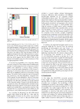
Figure 8. BMSCs proliferation results of the composite tablets for 1, 3,
and 5 days. that the bioactivity and osseointegration of samples are
improved.
to the crystal planes of (1 1 0), (1 1 3), (2 0 0), and (2 1 1), In addition, the improved biological activity of the
respectively. With the increase in the weight ratio of AKM, composite could also be observed from the evolving
the peaks belonging to AKM appeared more apparent, and morphology of hydroxyapatite over time (Figure 11).
the diffraction intensity of PEEK gradually decreased. The For pure PEEK, only a few microparticles adhered to
peak at 28.82° became sharpened because of the increasing the material surface with a time extension to 7 days,
intensity of the peak at 28.92° that belongs to AKM. No indicating weak bioactivity. By contrast, a large number
significant changes in the peak width and position occurred. of microparticles were attached to the surface of
Meanwhile, no new characteristic peaks appeared after 15 wt% AKM/PEEK composite on the first day. It was
sintering, which suggests that the AKM is stable in the accompanied by the formation of petal-like hydroxyapatite
process. It is therefore conducive to the maintenance of the with a size of fewer than 2 μm. With the extension of time
biological properties of AKM. to the seventh day, the petal-like hydroxyapatite grew to
To test the biocompatibility of the composites, BMSC a larger size exceeding 6 μm. This is mainly because the
cells were cultured for 5 days on the composite tablets in the Si ions released by AKM in the SBF solution promoted
[48]
medium. A control group was set for comparison, in which the formation and growth of hydroxyapatite . On the
BMSCs were cultured in a medium without additives. 14th day, the composite was covered with a dense network
Figure 8 shows the results obtained by a CCK-8 assay. In layer of hydroxyapatite. These results indicate that the
all groups, the absorbance values increased with time, and biological activity of the material is greatly improved with
there was no significant difference in absorbance among the incorporation of AKM.
groups. This means that the composite materials have good
cell proliferation and biocompatibility compared with 4. Conclusion
PEEK material. The cells cultured for 1 day and 5 days were In this study, the AKM/PEEK composite powders
dyed and observed by confocal microscopy (Figure 9). In in the weight ratio from 0 to 15 wt% were prepared,
all groups, the cells were uniformly and densely attached and the effects of AKM on mechanical and biological
to the composite surface. Compared with the first day, the properties were investigated. The morphology, melting/
cytoskeletons of the cells (schematized by red color) on the crystallization properties, and thermal stability of the
fifth day show obvious growth, which proves that the cells composite powders were tested to evaluate the HT-LPBF
seeded on the tablets have a good growth condition. This processability. The results suggested that the tiny AKM
indicates that the composites have positive effects on cell particles are uniformly dispersed in the composite. The
proliferation, which demonstrates good biocompatibility.
composition of AKM has little effect on the sintering
For evaluating the bioactivity, the HT-LPBF-printed window but can delay the peak crystallization temperature
samples were soaked in the SBF for 7 days, and the to reduce part warping. The melting enthalpy decreases
hydroxyapatite formation on the surface was observed with the increase of AKM content, which may lead to a
by the SEM (Figures 10 and 11). The pure PEEK sample higher surface temperature during the HT-LPBF process
Volume 9 Issue 3 (2023) 154 https://doi.org/10.18063/ijb.699