
Page 17 - IJB-9-3
P. 17
International Journal of Bioprinting Bioprinting of PDAC microtissues for drug screening
A
B C
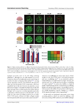
Figure 6. Drugs screening performed in different three-dimensional pancreatic ductal adenocarcinoma (PDAC) models after 7 days of culture.
(A) Representative fluorescent micrographs of PDAC models after incubation with gemcitabine ranging from 50 to 100 μM/mL for 72 h. Non-drug-treated
microtissues were observed as controls. Green channel: Calcein-AM. Red channel: PI. Scale bar = 200 μm. (B) Statistical plot of cell viabilities after treating
PDAC models with different concentrations of gemcitabine for 72 h. (C) Heat map representing the drug treatment results. Experimental values are
expressed in mean ± standard error, n = 3. Two-way ANOVA, *P < 0.05, **P < 0.01, ***P < 0.001, ****P < 0.0001.
hydrogel structures, serve as in vitro drug screening However, it is challenging to utilize some specific PDAC
platforms , although they are still restrained by several models in vitro due to their poor mechanical properties
[13]
[12]
limitations. Most cell-rich spheroids models mainly depend and slow crosslinking rate . As an alternative, gelatin-
on ultra-low plate or hanging drop method [20,36] , causing based hydrogels, as a hydrolysate of collagen matrices, are
tedious operations. Besides, the produced cell spheroids used because they exhibit good performance in building
[40]
often lack the biomimetic ECM components, which are 3D tumor models . In particular, GelMA is a kind of
considered one of the key elements in pancreatic cancer gelatin-based hydrogel, which is synthesized by modifying
microenvironment . Experiments employing certain cell- gelatin with methacrylated groups. The modified material,
[12]
laden hydrogel PDAC models based on the animal-derived which possesses the ability of rapid photocrosslinking and
Matrigel matrix are prone to yield unrepeatable results due the tunable mechanical capacity, emerges as a popular
[41]
to the batch-to-batch properties of the material . In view ECM-mimetic hydrogel in tissue engineering .
[39]
of the fact that a mass of collagen is present in the native In this work, we introduced a pancreatic cancer drug
PDAC ECM matrix, different cell-laden collagen models screening platform, which was made by utilizing a dot
have been produced for use in pancreatic cancer research . extrusion printing (DEP) system to print GelMA hydrogel
[21]
Volume 9 Issue 3 (2023) 9 https://doi.org/10.18063/ijb.v9i3.676