
Page 233 - IJB-9-3
P. 233
International Journal of Bioprinting Biomaterials for vascularized and innervated tissue regeneration
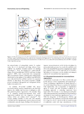
Figure 7. (A) The fabrication process of the 3D-bioprinted ossification center microenvironments biomimetic bone constructs. The bioinks were composed
of GelMA, AlgMA and NGF@Laponite. The constructs were crosslinked by UV light and Ca ions. (B) Mechanism of the 3D bioprinted constructs
2+
promoting the regeneration of bone defects[120]. Reprinted from Li W, Miao W, Liu Y, et al., Advanced Functional Materials, 2022, 202109871. Copyright
© 2021 John Wiley and Sons.
the mineralization of extracellular matrix. In another hypoxic microenvironment, which further stimulates the
study, Ha et al. developed dual-drug delivery bone neovascularization process. As a result, the scaffolds could
scaffolds via sacrificial templates-assisted 3D printing obviously upregulate the expression of angiogenesis-related
technology [118] . First, pro-angiogenesis small-molecule gene markers (such as VEGF and HIF-1α) of HUVECs,
drugs, dimethyloxalylglycine (DMOG) and bone forming and enhance its osteogenic activity, further confirming its
peptide-1 (BFP), were incorporated into mesoporous potential for vascularized bone regeneration.
silica nanoparticles (MSNs). Subsequently, DMOG/MSN
were loaded on the surface of the scaffolds, and BFP/MSN 4.3. 3D-printed biomaterials for innervated bone
were embedded into the scaffolds to achieve the sequential regeneration
delivery of dual drugs. 3D-printed composite scaffolds As previously mentioned, nerve fibers actively take part
[28]
possess satisfactory angiogenesis and osteogenesis ability in the process of bone regeneration and remodeling .
both in vitro and in vivo. Hence, simultaneous regeneration of neural elements in
the newly formed bone tissues is essential for functional
In addition, 3D-printed scaffolds with injury bone regeneration. The incorporation of neurotrophic
microenvironment response characteristics can obviously agents or neural cells into 3D-printed scaffolds is a
improve the viability and function of endogenous cells, promising approach to achieving innervated bone
thus promoting tissue regeneration. For example, Yang regeneration. For example, Li et al. developed ossification
et al. designed enzyme-functionalized bone tissue center microenvironment-mimicking 3D-bioprinted bone
regenerative scaffolds with the integration of glucose constructs for innervated bone regeneration (Figure 7) [120] .
oxidase (GOx) and catalase (CAT) enzymes [119] . The With the integration of NGF@Laponite (NGF@ Lap)
cascade catalytic reaction of GOx and CAT enzymes nanomaterials, the constructs continuously released NGF
could alleviate the hyperglycemic microenvironments and for a long time, which is similar to the microenvironment
continuously consume oxygen, leading to the formation of of ossification center with high concentration of NGF.
Volume 9 Issue 3 (2023) 225 https://doi.org/10.18063/ijb.706