
Page 232 - IJB-9-3
P. 232
International Journal of Bioprinting Biomaterials for vascularized and innervated tissue regeneration
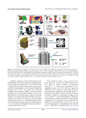
Figure 6. (A) The preparation and application of 3D-printed lotus root-like scaffolds for promoting bone regeneration and blood vessels infiltration [108] .
Reprinted from Feng C, Zhang W, Deng C, et al., Advanced Science, 2017, 4: 1700401. Copyright © 2017 John Wiley and Sons. (B) Schematic represen-
tation of microfluidic assisted-3D printed stimuli-responsive scaffolds with biomimetic hollow channels for promoting bone regeneration [110] . Reprinted
from Wang X, Yu Y, Yang C, et al., Advanced Functional Materials, 2021, 2105190. Copyright © 2021 John Wiley and Sons. (C) 3D printing of Haversian
bone-mimicking scaffolds for delivering BMSCs and HUVECs and promoting vascularized bone regeneration [111] . Reprinted from Zhang M, Lin R, Wang
X, et al., Science Advances, 2020, 6: eaaz6725. From ref. [111] licensed under Creative Commons Attribution 4.0 license.
In addition, designing a complex hierarchical structure Plenty of bioactive agents, such as growth factors,
scaffold similar to native bone tissues is a promising approach drugs, liposomes, enzymes and small molecules, have
to accelerating bone regeneration [100] . For instance, Zhang been shown to possess excellent pro-angiogenesis
et al. fabricated a Haversian canals-biomimetic bioceramic properties [103,112-117] . Hence, the incorporation of pro-
scaffold by using digital laser processing-based 3D printing angiogenesis agents into 3D-printed bone regenerative
technique (Figure 6C) [111] . The porosity and mechanical scaffolds has been regarded as a promising strategy for
strength of the biomimetic scaffolds could be accurately vascularized bone regeneration. For example, Han et al.
regulated by the custom design software. Moreover, the designed a lotus seedpod mimetic drug-loaded 3D-printed
hierarchical structure of biomimetic scaffolds can provide bioceramic scaffold for accelerating the healing process
a platform to modulate multicellular distributions and cell– of bone defects [116] . Deferoxamine@lipsome (DFO@lip)-
cell interactions. Besides, the Haversian bone-mimicking laden GelMA microsphere was fabricated via microfluidics
scaffolds could also serve as a multicellular delivery system technology and then integrated into 3D-printed β-TCP
for simultaneously inducing angiogenesis and osteogenesis scaffolds. The controlled release of DFO could not only
in vitro, and promoting vascularized bone regeneration in stimulate angiogenesis by upregulating hypoxia-inducible
a rabbit femoral defects model. factor 1 alpha (HIF-1α) expression but also promote
Volume 9 Issue 3 (2023) 224 https://doi.org/10.18063/ijb.706