
Page 227 - IJB-9-3
P. 227
International Journal of Bioprinting Biomaterials for vascularized and innervated tissue regeneration
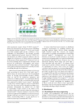
Figure 2. (A) The mechanism of peripheral nerves regulates the migration of mesenchymal stem cells and osteogenesis . Reprinted from Wang XD, Li
[29]
SY, Zhang SJ, et al., Theranostics, 2020, 10(11): 4839–4850. From ref. [29] licensed under Creative Commons Attribution 4.0 International License. (B) The
potential roles of neurotrophic factors in bone regeneration and remodeling . Reprinted from Su YW, Zhou XF, Foster BK, et al., Journal of Cellular
[10]
Physiology, 2018, 233(3): 2133–2145. Copyright © 2017 John Wiley and Sons.
with tropomyosin receptor kinase B (TrkB) receptors . In human body, blood vessel networks are distributed
[37]
BDNF also participates in the process of bone remodeling throughout tissues/organs for supplying nutrients and
by inducing osteoclast formation . Third, recent studies removing metabolic wastes. Nervous system extending
[38]
found that peripheral glial cell also contribute to bone highly branched neural fiber networks into targeted
metabolism and development . For example, paracrine tissues/organs to establish communication by transmitting
[39]
factors (platelet-derived growth factor-AA [PDGF-AA] electrical signals. . Developmental cues direct the
[44]
and oncostatin M [OSM]) secreted by Schwann cells can formation of neural and vascular networks in an ordered
regulate the cell behaviors of BSMCs and then participate manner with overlapping patterns to match the architectural
in the process of bone regeneration . Moreover, Cai et al. and functional demands of tissues. Moreover, due to the
[40]
found that Schwann cells co-cultured with osteoblast could anatomical similarity, blood vessels and nerves have a
promote the proliferation, differentiation, and calcium close interaction with each other . For instance, blood
[45]
nodules deposition activity of osteoblast . Taken together, vessels need to provide sufficient nutrients for supporting
[39]
neural system actively participates in bone development and the development of nerves, while nerves also regulate
remodeling through various pathways. vasodilation and vasoconstriction via transmitting signals .
[46]
Skeletal muscle tissue is the heaviest tissue in the human Moreover, previous studies also found that blood vessels and
[47]
body, accounting for about 45% of the body’s mass . nerve fibers are able to share same signals and receptors .
[41]
Muscle tissues are densely innervated with the peripheral For example, NGF, one of the most important neurotrophic
nerves by forming neuromuscular junctions (NMJs), factors for neuron maturation, has a positive effect on the
which are vital for the metabolism, maturation, and proliferation and migration activity of endothelial cells
[48,49]
contraction of skeletal muscle . It is well known that the through binding to its surface TrkA receptors . Similarly,
[42]
dynamic movement of body is realized through contractile VEGF could also promote the survival of neuron and axonal
[50]
force generated by skeletal muscle tissues . Specifically, outgrowth . Overall, considering the significant roles
[2]
somatic motor neurons trigger the release of acetylcholine of blood vessels and nerve fibers in tissue regeneration,
(Ach) from the axon terminal through generating action developing novel neurovascularized biomaterials based on
potentials. Subsequently, the released Ach can bind 3D printing technology is highly demanded.
to acetylcholine receptor (AchR) on the surface of the 3. Skin tissues
myofiber at NMJs, and eventually initiate the contraction of
skeletal muscle . However, volumetric muscle loss (VML) 3.1. 3D printing for skin tissue engineering
[43]
is always accompanied by significant motoneuron axotomy At present, auto-transplantation still remains the best
damage, resulting in permanent functional impairment . strategy in clinical setting for treating severe and critical-size
[2]
Hence, reconstructing NJMs is of great significance to skin defects, in which skin tissue is harvested from one part of
restore its physiological functions after VML. the body and subsequently grafted to injury site in the same
Volume 9 Issue 3 (2023) 219 https://doi.org/10.18063/ijb.706