
Page 228 - IJB-9-3
P. 228
International Journal of Bioprinting Biomaterials for vascularized and innervated tissue regeneration
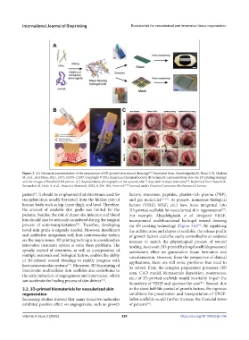
Figure 3. (A) Schematic representation of the preparation of 3D-printed skin wound dressings . Reprinted from Alizadehgiashi M, Nemr C R, Chekini
[64]
M, et al., ACS Nano, 2021, 15(7): 12375–12387, Copyright © 2021 American Chemical Society. (B) Schematic representation of in-situ 3D printing strategy
and the images of handheld 3D printer. (C) Representative photographs of the wounds after 7 days and 14 days treatments . Reprinted from Nuutila K,
[69]
Samandari M, Endo Y, et al., Bioactive Materials, 2022, 8: 296–308, From ref. licensed under Creative Commons Attribution 4.0 license.
[69]
patient . It should be emphasized that skin tissues used for factors, exosomes, peptides, platelet-rich plasma (PRP),
[1]
transplantation usually harvested from the hidden part of and gas molecules [53-62] . At present, numerous biological
human body, such as hip, inner thigh, and head. Therefore, factors (VEGF, bFGF, etc.) have been integrated into
the amount of available skin grafts was limited for the 3D-printed scaffolds for vascularized skin regeneration .
[63]
patients. Besides, the risk of donor site infection and blood For example, Alizadehgiashi et al. designed VEGF-
loss should also be seriously considered during the surgical incorporated multifunctional hydrogel wound dressing
[51]
process of auto-transplantation . Therefore, developing via 3D printing technology (Figure 3A) . By regulating
[64]
novel skin grafts is urgently needed. However, insufficient the architectures and shapes of scaffolds, the release profile
and ineffective integration with host neurovascular system of growth factors could be easily controlled in an ordered
are the major issues. 3D printing technique is considered an manner to match the physiological process of wound
innovative treatment option to solve these problems. The healing. As a result, 3D-printed hydrogel scaffolds possessed
specific control of structures, as well as incorporation of satisfactory effect on granulation tissue formation and
multiple materials and biological factors, enables the ability vascularization. However, from the perspective of clinical
of 3D-printed wound dressings to rapidly integrate with applications, there are still some problems that need to
host neurovascular systems . Moreover, 3D bioprinting of be solved. First, the complex preparation processes (3D
[14]
biomimetic multicellular skin scaffolds also contributes to scan, CAD model, biomaterials deposition, postprocess,
the early induction of angiogenesis and innervation, which etc.) of 3D-printed scaffolds would inevitably impair the
can accelerate the healing process of skin defects .
[52]
bioactivity of VEGF and increase the cost . Second, due
[65]
3.2. 3D-printed biomaterials for vascularized skin to the short half-life period of growth factors, the rigorous
regeneration conditions for preservation and transportation of VEGF-
Increasing studies showed that many bioactive molecules laden scaffolds would further increase the financial stress
exhibited positive effect on angiogenesis, such as growth of patients .
[66]
Volume 9 Issue 3 (2023) 220 https://doi.org/10.18063/ijb.706