
Page 300 - IJB-9-3
P. 300
International Journal of Bioprinting 3D bioprinting as a prospective therapeutic strategy for LSCD
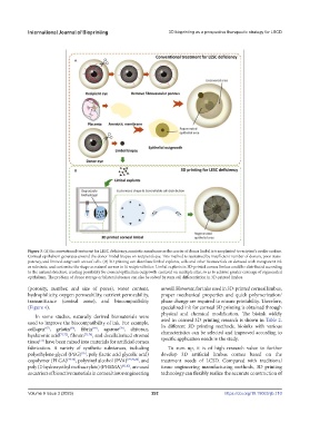
Figure 3. (A) In conventional treatment for LESC deficiency, amniotic membrane as the carrier of donor limbal is transplanted to recipient’s ocular surface.
Corneal epithelium generates around the donor limbal biopsy on recipient’s eye. This method is restrained by insufficient number of donors, poor trans-
parency, and limited outgrowth area of cells. (B) 3D printing can distribute limbal explants, cells and other biomaterials on demand with transparent ink
as substrate, and customize the shape as natural cornea to fit recipient better. Limbal explants in 3D-printed cornea limbus could be distributed according
to the natural structure, creating possibility for corneal epithelium outgrowth centered on multiple sites, so as to achieve greater coverage of regenerated
epithelium. The problem of donor storage or bilateral absence can also be solved by stem cell differentiation in 3D-printed limbus.
(porosity, number, and size of pores), water content, as well. However, for inks used in 3D-printed corneal limbus,
hydrophilicity, oxygen permeability, nutrient permeability, proper mechanical properties and quick polymerization/
transmittance (central zone), and biocompatibility phase change are required to ensure printability. Therefore,
(Figure 4). specialized ink for corneal 3D printing is obtained through
physical and chemical modification. The bioink widely
In some studies, naturally derived biomaterials were
used to improve the biocompatibility of ink. For example, used in corneal 3D printing research is shown in Table 2.
In different 3D printing methods, bioinks with various
[68]
[67]
[69]
collagen , gelatin , fibrin , agarose , chitosan, characteristics can be selected and improved according to
[70]
hyaluronic acid [71,72] , fibroin [73,74] , and decellularized stromal specific application needs in the study.
[75]
tissue have been mixed into materials for artificial cornea
fabrication. A variety of synthetic substances, including To sum up, it is of high research value to further
polyethylene glycol (PEG) , poly (lactic acid glycolic acid) develop 3D artificial limbus cornea based on the
[76]
copolymer (PLGA) [77,78] , polyvinyl alcohol (PVA) [77,79,80] , and treatment needs of LCSD. Compared with traditional
poly (2-hydroxyethyl methacrylate) (PHEMA) [81,82] , are used tissue engineering manufacturing methods, 3D printing
as carriers of bioactive materials in corneal tissue engineering technology can flexibly realize the accurate construction of
Volume 9 Issue 3 (2023) 292 https://doi.org/10.18063/ijb.710