Page 295 - IJB-9-3
P. 295
International Journal of Bioprinting                  3D bioprinting as a prospective therapeutic strategy for LSCD


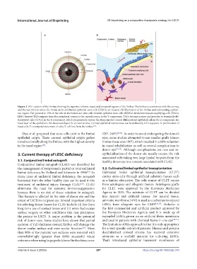
            Figure 2. (A) Location of the limbus showing the superior, inferior, nasal, and temporal regions of the limbus. The limbus is continuous with the cornea,
            and the red circle is where the limbal niche and limbal epithelial stem cells (LESCs) are located. (B) Illustration of the limbus and surrounding epitheli-
            um region. Five percent to 15% of the cells in the limbus are stem cells. Limbal epithelial stem cells (LESCs) divide into transit-amplifying cells (TACs).
            LESC-derived TACs migrate from the peripheral cornea to the central cornea in the Y component. TACs become mature and give rise to terminally dif-
            ferentiated cells (TDCs) in the X component, which progressively replace the desquamated corneal differentiated epithelial cells in the Z component. Ep:
            basal layer of the epithelium; BL: Bowman’s layer; St: corneal stroma. Corneal epithelial maintenance can be defined by XYZ equation. X: proliferation of
            basal cells, Y: centripetal movement of cells, Z: cell loss from the surface .
                                                         [14]
               Dua et al. proposed that stem cells exist in the limbal   120°–240°) [22,23] . In order to avoid endangering the donor’s
            epithelial crypts. These corneal epithelial crypts gather   eyes, some studies attempted to use smaller grafts (donor
            nondirectionally along the limbus, with the highest density   limbus tissue area <90°), which resulted in a 60% reduction
                           [18]
            in the nasal region .                              in visual rehabilitation as well as several complications in
                                                               donor eye [24,25] . Although complications are rare and re-
            3. Current therapy of LESC deficiency              epithelialization of the donor site usually occurs, the risk
                                                               associated with taking two large limbal biopsies from the
            3.1. Conjunctival limbal autograft                 healthy donor eye is a concern associated with CLAU.
            Conjunctival limbal autograft (CLAU) was described for
            the management of symptomatic partial or total unilateral   3.2. Cultivated limbal epithelial transplantation
            limbal deficiency by Holland and Schwartz in 1996 . In   Cultivated limbal epithelial transplantation (CLET)
                                                     [19]
            many  cases  of  unilateral  limbal  deficiency,  the  autograft   carries stem cells through artificial substrate tissues such
            harvested from the other healthy eyes can be used in the   as a limbus alternative. The cells source of CLET comes
            treatment of unilateral injury through CLAU . CLAU   from autologous and allogenic tissues. Autologous grafts
                                                  [20]
            eliminates  the  need  for  systemic  immunosuppression   for CLET were approved by the European Medicines
            because  there  is  no  risk  of  tissue  rejection  in  autograft.   Agency in 2015. The substrate of CLET can be divided
            This therapy is affected by the state of donor eye and the   into  natural  and  artificial  tissues.  For  natural  tissue,
            extent of LSCD in recipient eye. Several important criteria   amniotic membrane (AM) is used as a substrate to expand
            for selecting donor tissues for CLAU include: (i) free from   LESCs from allogenic eyes for CLET [26,27] . Holoclar is
            long-term use of contact lenses and (ii) never had ocular   the first commercial and artificial product approved by
            surface surgery or other conditions that may predispose   the  European  Medicines  Agency,  and  it  is  made  up  of
            the patient to LSCD. A major problem is the potential   expanded LESCs grown on an artificial fibrin membrane
            risk of donor eyes. Some studies have shown that partial   and used in patients with chemical burns in one eye [28-31] .
            excision of full-thickness corneal limbus will damage the   The limitation of this approach is that it is only appropriate
            donor ocular surface and even ocular function . More   for a very specific cohort of patients. Human and porcine
                                                   [21]
            than 90% of the patients’ eye surfaces were restored with   decellularized corneal stroma has received extensive
            overwhelmingly (greater than 80%) successful visual   attention  as  a  scaffold  material  for  culturing  LESCs.
            outcomes when using large grafts (donor limbus tissue area:   Thai’s introduced epithelial basement membranes of


            Volume 9 Issue 3 (2023)                        287                          https://doi.org/10.18063/ijb.710

