
Page 391 - IJB-9-3
P. 391
International Journal of Bioprinting Bioprinting of a multicellular model
supernatant, the tissue was washed with PBS for six times. 2.11. Pharmacodynamic evaluation of antitumor
Diluted DAPI solution (1:10000, Sigma) was added for drugs
staining for 5 min at room temperature in the dark. The On the 7 day, 3D bioprinted tissue was used for the
th
supernatant was discarded, and the tissue was washed antitumor drug sensitivity test. First, two groups were set:
with PBS for five times. A laser confocal microscope was a 3D printing-S and a 2D culture group. The sensitivity
used for observation. The wavelength of the laser confocal of the two drug screening models to fluorouracil (5-FU),
excitation light was selected according to the fluorescent oxaliplatin, irinotecan, and other common antitumor drugs
secondary antibody. in colorectal cancer was evaluated. The concentration
2.10. Transcriptome sequencing and biological gradients of 5-FU, oxaliplatin and irinotecan were set to 0, 0.1,
information analysis 1, 10, 50, and 100 µM in both groups. Cell proliferation was
measured using the CCK8 assay, and dose-response curves
In the 2D culture group, Trizol method was used for were plotted using GraphPad 9. The median inhibitory
extraction of RNA. In the 3D culture group and 3D concentrations of the three chemotherapeutic drugs in
printing-S group, the printing body was lysed to obtain the two groups were calculated based on these results. 3D
lysate, and the tumor cells were obtained by centrifugation. printing-M was treated with different concentrations of
The samples were collected after extraction using the antitumor drugs (0, 0.1, 1, 10, 50, 100, 200, and 500 µM).
Trizol method. The 3D printing-M group adopted the 3D
concentric circle culture mode, and the outer interstitium 2.12. Statistical analysis
was directly removed under the microscope. The principle Data are expressed as mean ± standard deviation.
of removal is to completely remove the interstitium, and The independent t-test was used for comparison of
part of the tumor cells can be removed, if necessary, to independent samples between two groups, and SPSS 26.0
ensure that only tumor cells are left in the 3D bioprinted software (version 26.0; IBM Corp., Armonk, NY, USA) was
tissue, without interstitial cells. After that, the 3D used for statistical analysis. Statistical significance was set
bioprinted tissue was lysed, and the tumor cells were at P < 0.05. Data from at least three independent samples
obtained by centrifugation. This experiment focused on or triplicate experiments were used for all assays.
analyzing and exploring the effects of tumor-associated
macrophages M2 and endothelial cells on the gene 3. Results
expression of SW480 cells cultured in a 3D printing-M 3.1. Construction of 3D bioprinted multicellular
model. Total mRNA was isolated using the Trizol or model of colorectal cancer
RNeasy Mini kit (QIAGEN, Dusseldorf, Germany) and
reverse-transcribed using the Ambion kit (Austin, USA). Concentric axis dual-nozzle 3D bioprinting was used to
In vitro transcription was performed using 1 – 5 ng of construct a 3D multicellular model of colorectal cancer
cDNA as a template, and RNA was reverse-transcribed into in concentric circle model, with tumors in the inner
the sequencing library. Sequencing libraries were prepared ring and tumor stromal cells in the outer ring. The final
using the NEBNext UltraTM RNA Library Preparation Kit
(Illumina). The sequencing library was then sequenced
on the Illumina HiSeq platform to generate 125/150 bp
peer reads. Differentially expressed genes (DEGs) were
analyzed using the DeSeq2 software package. Genes with
P < 0.05 adjusted for DESeq2 were assigned to DEGs. The
clustering analyzer R package was used to implement the
Gene Ontology (GO) and Kyoto Encyclopedia of Genes
and Genomes (KEGG) enrichment analyses. GO terms
and KEGG pathways with P<0.05 after correction were
considered to be significantly enriched by DEGs. The
protein-protein interaction (PPI) network of DEGs from
the STRING database was obtained, and a confidence
of 0.400 was selected (version 11.0, https://string-db.
org/). Cytoscape3.6.1 software was used to construct a
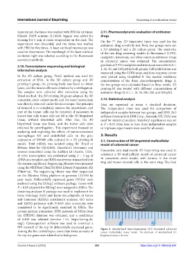
PPI network of the top 30 differentially expressed genes. Figure 2. Standardized three-dimensional (3D) bioprinted colorectal
Among the five central types, more than twice as many of cancer multicellular tissue model. The diameter of standardized 3D
the top ten genes were identified as hub genes. bioprinted tissue is 10 mm.
Volume 9 Issue 3 (2023) 383 https://doi.org/10.18063/ijb.694