
Page 392 - IJB-9-3
P. 392
International Journal of Bioprinting Bioprinting of a multicellular model
concentration of interstitial cells in the bio-ink was The survival of SW480 cells was observed using confocal
1.5 × 10 /mL. The diameter of the inner ring of the 3D microscopy. Green indicates live cell, and red indicates
6
printing-M was 7 mm, the diameter of the outer ring was cell death. As can be clearly seen from the images, a high
10 mm, and the thickness was 8 mm (Figure 2). Extrusion activity of >90% was maintained by day 10 (Figure 4).
3D bioprinting is used to construct stable 3D bioprinted The viability of macrophage and HUVEC cells in
models of colorectal cancer in high throughput. 3D hydrogel structure after 3D printing is shown in
Figure S1. Ki67 immunofluorescence was used to observe
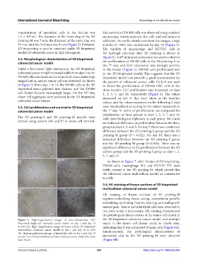
3.2. Morphological characteristics of 3D bioprinted the proliferation of SW480 cells in the 3D printing-S on
colorectal cancer model
th
the 7 day, and Ki67 expression was strongly positive
Under a low-power light microscope, the 3D bioprinted in the model (Figure 5). SW480 cells proliferated well
colorectal cancer model remained stable from days 1 to 10. in the 3D bioprinted model. This suggests that the 3D
SW480 cells were stable in the bioprinted tissue under high bioprinted model can provide a good environment for
magnification, and no tumor cell was observed. As shown the growth of colorectal cancer cells. CCK-8 was used
in Figure 3, from days 1 to 10, the SW480 cells in the 3D to detect the proliferation of SW480 CRC cells in the
bioprinted tissue gathered into clusters, and the SW480 three models. Cell proliferation was measured on days
cell clusters became increasingly larger. On the 10 day, 1, 3, 5, 7, and 10, respectively (Figure 6). The values
th
dense cell aggregates were scattered in the 3D bioprinted measured on the 1 day were taken as the baseline
st
colorectal cancer tissues. values, and the values measured on the following 4 days
3.3. Cell proliferation and survival in 3D bioprinted were standardized according to the values measured on
st
colorectal cancer model the 1 day. In terms of proliferation, we compared the
proliferation of three groups at days 1, 3, 5, 7, and 10,
The 3D printing-S and 3D printing-M models were with three biological replicates in each group. We found
stained using calcein AM and PI to assess cell survival. no statistical difference in proliferation between the three
groups at days 1, 3, and 5. On day 7, there was a statistical
difference between the 3D printing-S group and the 3D
A
printing-M group (P = 0.022). On day 10, there was a
statistical difference between the 3D printing-S group
and the 3D printing-M group (P=0.034). There was no
significant difference in the proliferation between the 3D
culture group and the 3D printing-S group at days 1, 3,
5, 7, and 10.
B As shown in Figure 7, after 10 days of 3D bioprinting,
SW480 cells, macrophage M2, and HUVEC-T/T were
stably present in the 3D printing-M, which proved that
the colorectal cancer multicellular model we constructed
is stable.
3.4. HE staining of frozen sections of 3D bioprinted
multicellular colorectal cancer model
C
HE staining of frozen sections of 3D printing-M
requires embedding, freeze curing, conventional paraffin
embedding, sectioning, fixation, staining, and sealing with
neutral gum. Tumor and interstitial cells were observed in
two parts under a microscope. HE staining characterized
the pathological characteristics of the tumor cell cluster in
Figure 3. High-magnification images of three-dimensional (3D) the 3D bioprinted colorectal cancer model, and multiple
bioprinted single-cell colorectal cancer model on day 1 and day 10 nuclei in the tumor cell cluster could be clearly seen,
in vitro (A). High-magnification images of tumor cells in 3D bioprinted indicating that it was composed of many cells (Figure 8A).
multicellular colorectal cancer model on day 1 and day 10 in vitro Simultaneously, the pathological characteristics of
(B). High-magnification images of interstitial cells on day 1 and day 10
in vitro in a 3D bioprinted multicellular colorectal cancer model (C). Scale interstitial cells in the 3D printing-M were observed
bars: 40 µm. (Figure 8B).
Volume 9 Issue 3 (2023) 384 https://doi.org/10.18063/ijb.694