
Page 389 - IJB-9-3
P. 389
International Journal of Bioprinting Bioprinting of a multicellular model
a 3D bioprinted single-cell model of colorectal cancer SW480 single-cell model was prepared by layer-by-layer
using SW480 cells as colorectal cancer seed cells and extrusion in a 3D bioprinter [24-26] . The print was immersed
gelatin/sodium alginate as bio-ink. The 3D bioprinted in a 100 mM calcium chloride solution for 3 min, and then
single-cell model was compared with the 2D and 3D 3 mL H-DMEM medium was added. The medium was
cultures. The differences in the morphology and biological changed every 2 days.
characteristics of the three culture models were evaluated.
We constructed a multicellular model for colorectal cancer 2.3. Construction of multicellular model for
drug screening using 3D bioprinting technology, developed colorectal cancer using dual-nozzle 3D bioprinter
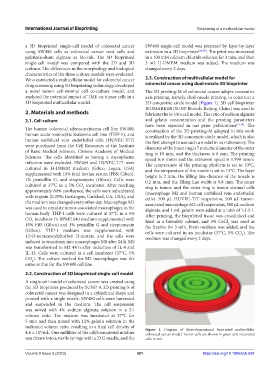
a novel tumor cell-stromal cell co-culture model, and The 3D printing-M of colorectal cancer adopts concentric
analyzed the potential impact of TME on tumor cells in a axis printing, namely, dual-nozzle printing, to construct a
3D bioprinted multicellular model. 3D concentric circle model (Figure 1). 3D cell bioprinter
BIOMARKER (SUNP Biotech, Beijing, China) was used to
2. Materials and methods fabricate the in vitro cell model. The ratio of sodium alginate
2.1. Cell culture and gelatin concentration and the printing parameters
have been reported in our prior publications [17,19] . The
The human colorectal adenocarcinoma cell line SW480, construction of the 3D printing-M adopted in this work
human acute monocytic leukemia cell line (THP-1), and is realized by the 3D concentric circle model, which is also
human umbilical vein endothelial cells (HUVEC-T/T) the first attempt to use such a model in our laboratory. The
were purchased from the Cell Resources of the Institute diameter of the inner ring is 7 mm, the diameter of the outer
of Basic Medical Sciences, Chinese Academy of Medical ring is 10 mm, and the thickness is 8 mm. The printing
Sciences. The cells identified as having a mycoplasma speed is 6 mm/s and the extrusion speed is 1.599 mm/s.
infection were excluded. SW480 and HUVEC-T/T were The temperature of the printing platform is set to 10°C,
cultured in H-DMEM medium (Gibco, Logan, USA) and the temperature of the nozzle is set to 15°C. The layer
supplemented with 10% fetal bovine serum (FBS; Gibco), height is 2 mm, the filling line distance of the nozzle is
1% penicillin G, and streptomycin (Gibco). Cells were 0.2 mm, and the filling line width is 0.8 mm. The inner
cultured at 37°C in a 5% CO incubator. After reaching ring is tumor, and the outer ring is tumor stromal cells
2
approximately 80% confluence, the cells were subcultured (macrophage M2 and human umbilical vein endothelial
with trypsin (0.25%; Invitrogen, Carlsbad, CA, USA), and cells). 500 µL HUVEC-T/T suspension, 500 µL tumor-
the medium was changed every other day. Macrophage M2 associated macrophage M2 cell suspension, 500 µL sodium
was used to simulate tumor-associated macrophages in the alginate, and 1 mL gelatin were added at a ratio of 1:1:1:1.
human body. THP-1 cells were cultured at 37°C in a 5% After printing, the bioprinted tissue was crosslinked and
CO incubator in RPMI1640 medium supplemented with fixed in a biosafety cabinet, and 3% CaCl was used as
2
10% FBS (Gibco) and 1% penicillin G and streptomycin the fixative for 3 min. Fresh medium was added, and the
2
(Gibco). THP-1 medium was supplemented with cells were cultured in an incubator (37°C, 5% CO ). The
12-O-tetracanoylphorbol 13-acetate, and the cells were medium was changed every 2 days. 2
induced to transform into macrophages M0 after 24 h. M0
was transformed to M2 48 h after induction of IL-4 and
IL-13. Cells were cultured in a cell incubator (37°C, 5%
CO ). The culture method for M2 macrophages was the
2
same as that for the SW480 cell line.
2.2. Construction of 3D bioprinted single-cell model
A single-cell model of colorectal cancer was created using
the 3D bioprinter produced by SUNP. A 3D printing-S of
colorectal cancer was designed in a cylindrical shape and
printed with a single nozzle. SW480 cells were harvested
and suspended in the medium. The cell suspension
was mixed with 4% sodium alginate solution in a 2:1
volume ratio. The mixture was incubated at 37°C for
5 min and then mixed with 12% gelatin solution in the
indicated volume ratio, resulting in a final cell density of Figure 1. Diagram of three-dimensional bioprinted multicellular
4.8 × 10 /mL. One milliliter of the cell/biomaterial mixture colorectal cancer model. Tumor cells are shown in green and interstitial
6
was drawn into a sterile syringe with a 23 G needle, and the cells in red.
Volume 9 Issue 3 (2023) 381 https://doi.org/10.18063/ijb.694