
Page 436 - IJB-9-3
P. 436
International Journal of Bioprinting Gelatin-PVA crosslinked genipin bioinks for skin tissue engineering
leachate media. For each of the three biological samples 2.21. Statistical analysis
(n = 3), three technical replicates were conducted. A Nikon All experiments were repeated at least 3 times (n = 3), and
A1R-A1 live-cell imaging microscopy was used to obtain statistical analyses were performed at the significance level
pictures at 1-h intervals to estimate the wound-healing rate of P < 0.05 by one-way and two-way analysis of variance
as follows:
(ANOVA) using GraphPad Prism (V9.0, GraphPad
Initialareaof Software Inc., San Diego, CA, USA).
thewound( µ m ) −
2
3. Results
Finalareaofthhe
3.1. Gross appearance
wound(µ m )
2
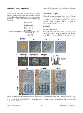
Healingprogress(%) = × 100 Figure 2A indicates that the digital photograph of the
Initialareaofthe 3D-bioprinted GPVA bioinks was printed with different
wound µ ( m ) layers (1 layer, 3 layers, and 5 layers) through extrusion-
2
A
C
B
D
Figure 2. Gross appearance of the 3D-printed hydrogels. (A) Autocad design in stl. file followed by different layers of printed bioinks (1 layer, 3 layers,
and 5 layers). (B) Optimization of printing temperature for the bioinks (under-gelation printed hydrogel (25 ± 2°C), optimum gelation printed hydrogel
(23 ± 2°C), over-gelation printed hydrogel (19 ± 2°C). (C) Width evaluation of 3D-printed hydrogels. (D) Gross appearance of NC and GNP hydrogels
for GE, GPVA3, and GPVA5.
Volume 9 Issue 3 (2023) 428 https://doi.org/10.18063/ijb.677