
Page 437 - IJB-9-3
P. 437
International Journal of Bioprinting Gelatin-PVA crosslinked genipin bioinks for skin tissue engineering
based bioprinting. The top view images demonstrate that characterized based on biodegradation rate, contact
the hydrogels have a high printing precision to deposit angle, swelling ratio, WVTR, degree of crosslinking, and
bioinks up to 5 layers with better shape fidelity. Moreover, antioxidant properties. The physicochemical analysis
the printability assessment of the bioinks was evaluated was evaluated for GNP and NC hydrogels, as shown
against different printing temperatures (19 ± 2°C, 23 ± in Figure 3. Hydrogels should possess multifunctional
2°C, and 25 ± 2°C) as shown in Figure 2B. The printed properties to accelerate wound healing. In wound healing
hydrogels’ stability and shape fidelity decreased and applications, cellular skin replacements are designed
slightly collapsed as temperature increased. However, with an appropriate biomaterial that can degrade while
the filaments printed at optimum temperature were promoting a faster wound healing time frame. Adding
thinner (0.1 ± 0.025 cm) than other filaments printed GNP as a natural crosslinker and PVA helps improve
at under- and over-gelation (0.37 ± 0.03 and 0.21 ± the stability and control the biodegradability of the
0.04 cm) as shown in Figure 2C. Next, six different types hydrogels, as stated in Figure 3A. The biodegradation
of bioinks formulations were printed using GE_NC, rate of GPVA5_GNP was found to be the slowest (0.018 ±
GPVA3_NC, GPVA5_NC, GE_GNP, GPVA3_GNP, and 0.08 mg/h) followed by GPVA3_GNP and GE_GNP (0.023
GPVA5_GNP through extrusion-based bioprinting, ± 0.21 mg/h and 0.062 ± 0.11 mg/h). However, all NC
respectively. The hydrogels in Figure 2C were printed hydrogels were totally degraded within 1 h. The common
with 5 layers of bioinks and were characterized by factor of such assessment is hydrophilicity, which is related
well-defined individual printheads, as the composite to the water contact angle and is reflected by the moisture
[30]
hydrogels’ grid is clearly seen at size 2.5 cm . Genipin content of the biomaterial . The water contact angle
2
(GNP) was used to construct and chemically crosslink values of the hydrogels are shown in Figure 3B. For GNP
gelatin-PVA hydrogels. Crosslinked hydrogels appeared hydrogels, GE_GNP has the lowest contact angle (42.93 ±
bluish in color while non-crosslinked hydrogels 3.2°), followed by GPVA3_GNP and GPVA5_GNP (49.36
appeared colorless after printing. Figure 2D shows the ± 2.1° and 50.2 ± 1.2°). Besides, NC hydrogels GE_NC,
gross appearance of the 3D-bioprinted hydrogels for GPVA3_NC, and GPVA5_NC have slightly larger contact
non-crosslink and crosslinked hydrogels. angles compared to GNP hydrogels (44.8 ± 4.6°, 51.2±3.6°,
and 53.5 ± 4.0°), respectively.
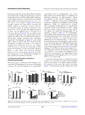
3.2. Physical and antioxidant properties of In wound healing applications, hydrogel was selected
3D-bioprinted hydrogel because its 3D networks are made up of hydrophilic
This study focused on fabricating GPVA hydrogels with polymers that expand in an aqueous solution and have
crosslinked GNP through an extrusion-based bioprinting the capacity to retain some water without dissolving.
technique. The fabricated hydrogels were systematically Figure 3C demonstrates that the swelling ratio of the
A B C D
E
F G
Figure 3. The physical and antioxidant analysis of 3D-printed hydrogels: (A) biodegradation rate, (B) contact angle, (C) swelling ratio, (D) water vapor
transmission rate (WVTR), (E) degree of crosslinking, (F) DPPH assay, and (G) ABTS assay. *P < 0.05.
Volume 9 Issue 3 (2023) 429 https://doi.org/10.18063/ijb.677