
Page 442 - IJB-9-3
P. 442
International Journal of Bioprinting Gelatin-PVA crosslinked genipin bioinks for skin tissue engineering
A B
C
D
E
F
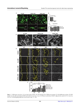
Figure 7. Cell-bioinks interaction. (A) Live/dead assay of HDFs. (B) Cell viability of the HDFs post-printing. (C) Cell proliferation activity. (D) SEM
images show the cell morphology of HDFs in the bioink (scale bar, 10 µm). (E) Wound scratch assay of HDFs using leachate media for 72 h (scale bar,
100 µm). (F) Healing progression for wound scratch assay. *P < 0.05.
Volume 9 Issue 3 (2023) 434 https://doi.org/10.18063/ijb.677