
Page 48 - IJB-9-3
P. 48
International Journal of Bioprinting Curved cell-guided structures printed by FDM
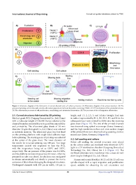
Figure 1. Schematic diagram of the fabrication of curved channels and cell culture processes. (A) Illustrative diagram of the process markers. (B) The
process of printing curved channels onto the siliconized glass sheets by fused deposition modeling (FDM). (C) UV sterilization of the printed structures.
(D) The observation processes of cell proliferation, morphology, and orientation observation. (E) The processes of the cell migration observation.
2.1. Curved structures fabricated by 3D printing height, and 1.5, 2, 2.5, 3, and infinite (straight line) mm
Medical-grade PCL (Daigang Biomaterial Co., Ltd, China) in radius (represented by R1.5, R2, R2.5, R3, and SL in the
with a molecular weight of 150,000 Da was selected as the subsequent text) were printed by FDM onto the receiving
channel boundary material due to its good biocompatibility glass sheet (Figure 1B). The size of the printed channels
and printability. The siliconized glass sheets of 24 mm was observed using optical microscope (Leica, Germany),
diameter (JingAn Biological Co., Ltd, China) were selected and the high-resolution surface and cross-section images
as substrate material. The siliconized glass was first fixed of the printed fibers were observed using scanning electron
on the printing platform with single-sided adhesive tape microscope (Zeiss, Germany) (Figure S1).
before printing. The starting point of printing was selected
at the center of the glass sheet. The inner diameter of 2.2. Cell seeding and culture
the needle for structure printing was 100 μm. Two-stage Before cell seeding, the printed structures were placed
temperature control was employed to heat the PCL, in the culture dishes and sterilized with ultraviolet (UV)
with the temperature being set as 120°C and 105°C, light in a UV sterilization chamber (Kangrong Biomedical
respectively. The air pressure of the printer was 0.6 MPa, Technology Co., Ltd, China) for 3 h (Figure 1C). The
and the platform movement speed was 0.8 mm/s. After the sterilized substrates were transferred into a clean six-well
pressured air inlet was closed, the platform was configured plate (JingAn Biological Co., Ltd, China).
to elevate automatically and slowly to prevent the inertia Human embryonic fibroblast M-22 cell (M-22 cell) was
extrusion of fibers from damaging the designed structures. spindle-shaped with a rapid migration and proliferation
Predesigned channels with 100 μm in width, 150 μm in speed, suitable for observing the cell orientation and
Volume 9 Issue 3 (2023) 40 https://doi.org/10.18063/ijb.681