
Page 62 - IJB-9-3
P. 62
International Journal of Bioprinting Peritoneal scaffolds for the peritoneal adhesion prevention
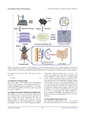
Scheme 1. Fabrication and mechanism of peritoneal scaffolds in the prevention of peritoneal adhesions. Step 1: scaffolds with different pore shapes were
constructed using MEW technology. Step 2: Peritoneal mesothelial cells were extracted from mice for in vitro expansion and culture. Step 3: Peritoneal me-
sothelial cells were cocultured with the scaffold. Step 4: Scaffolds loaded with peritoneal mesothelial cells were transplanted to prevent peritoneal adhesion.
the integral of the area under the tensile stress (τ)–strain streptomycin antibiotics, and cut into 1-mm pieces. The
(ε) curve. tissues were washed twice with PBS and digested with
trypsin (PB180225; Procell Life Science and Technology
U = ∫τdε (I) Co. Ltd., Wuhan, China) for 20 min in a water bath at 37°C.
2.4. Detection of contact angles The trypsin reaction was terminated by adding complete
The contact angles of PCL scaffolds were determined at culture medium of mouse peritoneal mesothelial cells. The
room temperature using a contact angle goniometer (SZ- cell suspension was gently resuspended by pipette until
CAMD33; Shanghai Sunzern Instrument Co. Ltd., China) there was no lumpy tissue, and then centrifuged at 200 × g
by the sessile drop method. The value of each sample was for 8 min after passing through a 100-μm mesh sieve. Cell
measured in triplicate. precipitate was retained, while supernatant was discarded.
The cells were resuspended with complete medium of
2.5. Isolation and culture of primary mesothelial cells mouse peritoneal mesothelial cell and inoculated in a
After killing the mice, they were soaked in 75% alcohol culture dish precoated with polylysine at 37°C in a 5% CO 2
for 5 min, moved to an ultraclean bench, and placed in incubator.
supine position during the experiment. The skin was
then cut along the midline of the abdomen, and the 2.6. Degradation experiment in vivo
peritoneal tissue was removed aseptically and immersed The peritoneal scaffolds measuring 0.5 × 0.5 cm were
in phosphate-buffered saline (PBS) containing penicillin- fixed onto the peritoneum of mice. At each predesignated
Volume 9 Issue 3 (2023) 54 https://doi.org/10.18063/ijb.682