
Page 66 - IJB-9-3
P. 66
International Journal of Bioprinting Peritoneal scaffolds for the peritoneal adhesion prevention
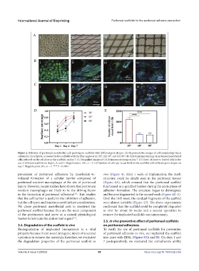
Figure 2. Behavior of peritoneal mesothelial cells growing on scaffolds with different pore shapes. (A) Representative images of cell morphology when
cultured in (i) cell plate, or loaded by the scaffolds with the fiber angles at (ii) 30°, (iii) 60°, and (iv) 90°. (B) CK18 immunostaining of peritoneal mesothelial
cells cultured on the cell plate or the scaffolds on day 7. (C) Magnified images of CK18 immunostaining on day 7. (D) Ratio of pores to loaded cells in the
use of different scaffolds on days 1, 4, and 7. Magnification: 20×, n = 3. (E) Number of cells per visual field on the scaffolds with different pore shapes on
day 7. Magnification: 20×, n = 3. ****P < 0.0001.
prevention of peritoneal adhesions by interleukin-4c- vivo (Figure 4). After 1 week of implantation, the mesh
induced formation of a cellular barrier composed of structure could be clearly seen in the peritoneal tissues
peritoneal resident macrophages at the site of peritoneal (Figure 4A), which ensured that the peritoneal scaffold
injury. However, recent studies have shown that peritoneal functioned as a qualified barrier during the acute phase of
resident macrophages are likely to be the driving factor adhesion formation. The structure began to disintegrate
in the formation of peritoneal adhesions . This implies and became fragmented in the second week (Figure 4B–E).
[26]
that the cell barrier is useful in the inhibition of adhesion, Until the 16th week, the residual fragments of the scaffold
but the cell types and functions need further consideration. were almost invisible (Figure 4F). The above experiments
We chose peritoneal mesothelial cells to construct the confirmed that the scaffold could be completely degraded
peritoneal scaffold because they are the main component in vivo for about 16 weeks and a second operation to
of the peritoneum and serve as a natural physiological remove the implanted scaffolds was unnecessary.
barrier to lubricate the abdominal organs .
[15]
3.5. In vivo preventive effect of peritoneal scaffolds
3.4. Degradation of the scaffold in vivo on peritoneal adhesions
Biodegradation of implanted biomaterials is a vital To verify the use of peritoneal scaffolds for prevention
property because it can avoid iatrogenic injury of a second of peritoneal adhesion in vivo, we implanted the scaffold
operation to remove the materials; therefore, we evaluated into mice with IBMs (Figure S3A and B). On days 1 and
the degradation properties of the peritoneal scaffold in 7 postoperatively, we evaluated the antiadhesive ability
Volume 9 Issue 3 (2023) 58 https://doi.org/10.18063/ijb.682