
Page 68 - IJB-9-3
P. 68
International Journal of Bioprinting Peritoneal scaffolds for the peritoneal adhesion prevention
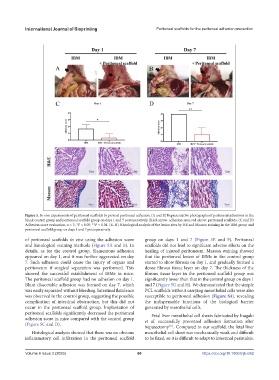
Figure 5. In vivo experiments of peritoneal scaffolds to prevent peritoneal adhesion. (A and B) Representative photographs of peritoneal adhesions in the
blank control group and peritoneal scaffold group on days 1 and 7 postoperatively. Black arrow: adhesion area; red arrow: peritoneal scaffolds. (C and D)
Adhesion score evaluation, n = 3. *P < 0.05; **P < 0.01. (E–H) Histological analysis of the lesion sites by HE and Masson staining in the IBM group and
peritoneal scaffold group on days 1 and 7 postoperatively.
of peritoneal scaffolds in vivo using the adhesion score group on days 1 and 7 (Figure 5E and F). Peritoneal
and histological staining methods (Figure 5A and B). In scaffolds did not lead to significant adverse effects on the
details, as for the control group, filamentous adhesion healing of injured peritoneum. Masson staining showed
appeared on day 1, and it was further aggravated on day that the peritoneal lesion of IBMs in the control group
7. Such adhesion could cause the injury of organs and started to show fibrosis on day 1, and gradually formed a
peritoneum if surgical separation was performed. This dense fibrous tissue layer on day 7. The thickness of the
showed the successful establishment of IBMs in mice. fibrous tissue layer in the peritoneal scaffold group was
The peritoneal scaffold group had no adhesion on day 1. significantly lower than that in the control group on days 1
Blunt dissectable adhesion was formed on day 7, which and 7 (Figure 5G and H). We demonstrated that the simple
was easily separated without bleeding. Intestinal flatulence PCL scaffolds without carrying mesothelial cells were also
was observed in the control group, suggesting the possible susceptible to peritoneal adhesion (Figure S4), revealing
complication of intestinal obstruction, but this did not the indispensable functions of the biological barrier
occur in the peritoneal scaffold group. Implantation of generated by mesothelial cells.
peritoneal scaffolds significantly decreased the peritoneal Fetal liver mesothelial cell sheets fabricated by Inagaki
adhesion score in mice compared with the control group et al. successfully prevented adhesion formation after
(Figure 5C and D).
hepatectomy . Compared to our scaffold, the fetal liver
[11]
Histological analysis showed that there was no obvious mesothelial cell sheet was mechanically weak and difficult
inflammatory cell infiltration in the peritoneal scaffold to be fixed, so it is difficult to adapt to intestinal peristalsis.
Volume 9 Issue 3 (2023) 60 https://doi.org/10.18063/ijb.682