
Page 69 - IJB-9-3
P. 69
International Journal of Bioprinting Peritoneal scaffolds for the peritoneal adhesion prevention
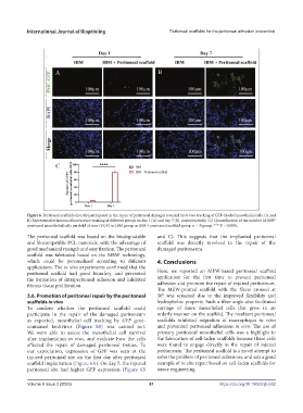
Figure 6. Peritoneal scaffolds directly participated in the repair of peritoneal damages revealed by in vivo tracking of GFP-labeled mesothelial cells. (A and
B) Representative immunofluorescence staining of different groups on day 1 (A) and day 7 (B), postoperatively. (C) Quantification of the number of GFP +
peritoneal mesothelial cells per field of view (FOV) in IBM group or IBM + peritoneal scaffold group. n = 3/group. ****P < 0.0001.
The peritoneal scaffold was based on the biodegradable and C). This suggests that the implanted peritoneal
and biocompatible PCL materials, with the advantage of scaffold was directly involved in the repair of the
good mechanical strength and easy fixation. The peritoneal damaged peritoneum.
scaffold was fabricated based on the MEW technology,
which could be personalized according to different 4. Conclusions
applications. The in vivo experiments confirmed that the
peritoneal scaffold had good biosafety, and prevented Here, we reported an MEW-based peritoneal scaffold
the formation of intraperitoneal adhesion and inhibited application for the first time to prevent peritoneal
fibrous tissue proliferation. adhesion and promote the repair of injured peritoneum.
The MEW-printed scaffold with the fibers crossed at
3.6. Promotion of peritoneal repair by the peritoneal 30° was screened due to the improved flexibility and
scaffolds in vivo hydrophobic property. Such a fiber angle also facilitated
To confirm whether the peritoneal scaffold could carriage of more mesothelial cells that grew in an
participate in the repair of the damaged peritoneum orderly manner on the scaffold. The resultant peritoneal
as expected, mesothelial cell tracking by GFP gene- scaffolds inhibited migration of macrophages in vitro
contained lentivirus (Figure S5) was carried out. and prevented peritoneal adhesions in vivo. The use of
We were able to assess the mesothelial cell survival primary peritoneal mesothelial cells was a highlight in
after implantation in vivo, and evaluate how the cells the fabrication of cell-laden scaffolds because these cells
affected the repair of damaged peritoneal tissues. To were found to engage directly in the repair of injured
our expectation, expression of GFP was seen at the peritoneum. The peritoneal scaffold is a novel attempt to
injured peritoneal site on the first day after peritoneal solve the problem of peritoneal adhesions, and sets a good
scaffold implantation (Figure 6A). On day 7, the injured example of in situ repair based on cell-laden scaffolds for
peritoneal site had higher GFP expression (Figure 6B tissue engineering.
Volume 9 Issue 3 (2023) 61 https://doi.org/10.18063/ijb.682