
Page 12 - IJB-9-4
P. 12
International Journal of Bioprinting Biomimetic 3D printed glioma model
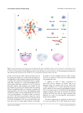
Figure 1. Diagram of glioma microenvironment and the bioprinted 3D models. (A) Diagram of glioma microenvironment, in which glioma cells are
wrapped with neurons . (B) The bioprinted glioma in vitro 3D model with glioma cells in the inner layer and neurons in the outer layer (G/N) (d = 8 mm,
[29]
D = 12 mm). (C) The bioprinted 3D model with glioma cells in the inner layer and no cell in the outer layer (i.e., G). (D) The bioprinted 3D model with no
cells in the inner layer and neurons in the outer layer (i.e., N). The dimensions of the three models are the same.
printer to form the print paths. After the parameter was set for culture in the same medium as neurons, which has been
for printing, the bioink was extruded and assembled in a proven to be suitable for GL261 cells. The culture medium
receiving mold. As the forming container for the model, the was changed every 48 h.
receiving mold consisted of hemispherical silicone negative In addition, in order to better understand the
mold with a diameter of 12 mm and hemispherical UV- interactions between neurons and glioma cells in the
cured positive mold with a diameter of 8 mm to match the designed model, two controls were set up, which were
fabrication method. After printing neuron bioink into the G and N conditions. For G control, the bioink used was
gap between the positive and negative mold, the resulting GL261 cell bioink in the inner layer and collagen (2 mg/mL,
external hemispherical shell was completely crosslinked by pH 7.4) in the outer layer. For N control, the bioink used was
being incubated at 37°C for 30 min. The preliminary model collagen (2 mg/mL, pH 7.4) in the inner layer and neuron
was taken out of the incubator and gently placed at the center bioink in the outer layer. The two types of models were
of the printing platform. The positive mold was removed fabricated by means of performing the aforementioned
and GL261 cell bioink was printed to shape the internal biofabrication method.
hemisphere, which was incubated at 37°C for another period
of 30 min. The glioma in vitro 3D model was formed, and For better representing the hierarchical structure of
then placed into non–tissue-culture-treated 24-well plates the constructed glioma 3D model, Trypan Blue solution
Volume 9 Issue 4 (2023) 4 https://doi.org/10.18063/ijb.715