
Page 17 - IJB-9-4
P. 17
International Journal of Bioprinting Biomimetic 3D printed glioma model
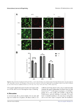
Figure 6. The survival rate of neurons in G/N and N after 1, 3, and 5 days of culture. (A) Confocal images of live/dead staining (green color: live cells; red
color: dead cells) of neurons. (B) Histograms of the survival rate of neurons in G/N and N. Significance was calculated using two-way ANOVA followed
by t-test. All error bars are standard deviation. ∗p < 0.05, ∗∗p < 0.01, ∗∗∗p < 0.001, and ∗∗∗∗p < 0.0001, respectively.
G/N outgrew significantly more neurites and longer axons scaffolds after being mixed evenly, which cannot precisely
in G/N than that found in N during the 5 days of culturing. represent the spatial relationship of different types of cells
found in vivo [30-32] . In this paper, we propose a novel bilayer
glioma 3D in vitro model and provide a step-by-step
4. Discussion biofabrication method utilizing a removable UV-cured
In conventional cell co-culture models, two or more cell mold to bioprint neurons in the outer layer and glioma
types are seeded onto 2D planar cell culture dishes or 3D cells in the inner layer. Compared with the traditional
Volume 9 Issue 4 (2023) 9 https://doi.org/10.18063/ijb.715