
Page 18 - IJB-9-4
P. 18
International Journal of Bioprinting Biomimetic 3D printed glioma model
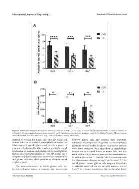
Figure 7. Neurite characteristics of individual neurons in G/N and N after 1, 3, and 5 days of culture. (A) Number of neurites of individual neuron in
G/N and N. (B) Axon length of individual neuron in G/N and N. Significance was calculated using two-way ANOVA followed by t-test. All error bars are
standard deviation. ∗p < 0.05, ∗∗p < 0.01, ∗∗∗p < 0.001, and ∗∗∗∗p < 0.0001, respectively.
method of seeding the mixed cells onto 2D planar cell between glioma cells and neurons have important
culture dishes or 3D scaffolds, this method can realize the influences for progression of glioma. In the bioprinted
fabrication of a spatially partitioned co-culture model of glioma in vitro 3D model, the glioma cells close to neurons
neurons and glioma cells, which can best mimic the spatial were found elongated with filopodium in morphology.
relationship of neurons and glioma cells in actual glioma Filopodium is a typical feature of neural cells, and this
tissues. The bioprinted glioma in vitro 3D model has a result indicates that neurons promote the transformation
complete and clear demarcation line between neurons area to neuron-like cells of GL261 cells, which is consistent with
and glioma cells area, which provides an adequate model the phenomenon observed in vivo and in vitro [16,33,34] . In
[18]
of glioma tissue. actual glioma tissues, glioma cells outgrow filopodium
The microenvironment in which glioma cells live to establish electrical connection with neurons around
in natural human tissues is complex, and interactions them to enhance invasiveness. But on the other hand,
[18]
Volume 9 Issue 4 (2023) 10 https://doi.org/10.18063/ijb.715