
Page 142 - IJB-9-5
P. 142
International Journal of Bioprinting 3D printed PEEK scaffold mediates macrophages to affect osseointegration
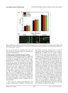
Figure 2. Proliferation and adhesion of cells on PEEK scaffolds with different pore sizes. (A) CCK-8 results of M1 macrophage cultured on PEEK scaffold
with different pore sizes (0, 200, and 400 μm) for 1, 4, and 7 days. (B) The fluorescence staining of macrophages co-cultured with scaffolds of different pore
sizes for 7 days (scale bar = 200 μm).
pore size can promote the anti-inflammatory effect of direct effect of paracrine secretions after co-culture of
macrophages and increase the release of osteogenesis and macrophages with scaffolds on the induction of osteogenic
angiogenesis-related factors. differentiation of BMSCs, the ALP activity of BMSCs
after 14 days of culture in CM was examined, as shown
3.3. Macrophage-mediated osteogenic activity in Figure 5B. The results showed that the ALP activity in
To evaluate the immunomodulatory effect of samples, the the PEEK group was relatively the lowest, while the ALP
supernatants from macrophage cultures were used as CM activity in the PEEK400 group was the highest, and there
to evaluate the osteogenic activity of BMSCs. The time- was a statistical difference between the two groups. The
based proliferation of BMSCs is shown in Figure 4. After results of ALP staining (Figure 5A) showed that the ALP
incubation for 1 day, no obvious difference was reported staining in the PEEK200 and PEEK400 scaffold groups
among the three groups. The PEEK200 and the PEEK400 was significantly deeper than that in the PEEK group after
showed higher proliferation rate in comparison with the BMSCs were induced and cultured for 14 days, and the
PEEK after 4 and 7 days of culture, and the rabbit BMSCs PEEK400 group had the most obvious staining results,
exhibited much greater proliferation on the PEEK400 which reconfirmed the results of ALP. Furthermore, the
group (p < 0.01). Figure 4B showed the cell morphology Alizarin red S staining results (Figure 5D) showed that
of rabbit BMSCs cultured for 7 days. It can be seen that with the increase of pore size, the staining of Alizarin red
the cells in the PEEK group grow mono-dispersedly, S gradually became intense, and the semi-quantitative
while the cells in the PEEK400 group are connected to results of Alizarin red S staining (Figure 5C) showed that
each other and spread more obviously, indicating that the the OD value at 600 nm of the PEEK400 scaffold group
paracrine secretions after co-culture of macrophages with was significantly higher. This indicates that the paracrine
macropore-sized scaffolds can promote the proliferation secretion of macrophages in PEEK400 group induces a
and adhesion of BMSCs, which is consistent with the higher level of extracellular mineralization of BMSCs,
results of CCK-8.
which also means that it has the strongest effect on
ALP is one of the commonly used indicators for the inducing osteogenic differentiation, which can be mutually
osteogenic differentiation of BMSCs. To examine the confirmed with the results of ALP activity.
Volume 9 Issue 5 (2023) 134 https://doi.org/10.18063/ijb.755