
Page 144 - IJB-9-5
P. 144
International Journal of Bioprinting 3D printed PEEK scaffold mediates macrophages to affect osseointegration
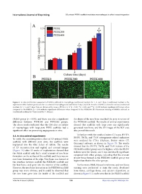
Figure 4. In vitro proliferation assessment of BMSCs cultured in macrophage-conditioned medium for 1, 4, and 7 days. Conditioned medium is the
supernatant of the medium produced after co-culture of macrophages and scaffolds for 3 days. (A) CCK-8 results of BMSCs cultured in various conditioned
medium for 1, 4, and 7 days, Data are shown as the mean ± SD for n ≥ 3; *p < 0.05, **p < 0.01, and ***p < 0.001 indicate significant differences when
compared to the PEEK0; p < 0.05 indicates significant differences when compared to the PEEK200. (B) Fluorescent staining of BMSCs cultured in
#
conditioned medium for 7 days (scale bar = 100 μm).
PEEK0 group (p < 0.05), and there was also a significant the shape of the new bone matched the pore structure of
difference between PEEK400 and PEEK200 groups. the PEEK400 scaffold. The results of animal experiments
The above results indicated that the CM after co-culture showed that scaffolds with large pore size significantly
of macrophages with large-pore PEEK scaffolds had a generated new bone, and the 3D images of the ROI area
significant effect on promoting angiogenesis in vitro. also proved this result.
3.5. In vivo animal experiments To further verify the results of micro-CT scans, BV/TV,
To verify the osseointegration effect of 3D-printed PEEK BS/BV, Tb.Th, and Tb.N osteogenesis-related indicators
scaffolds with different pore sizes, the scaffolds were were analyzed by CTAn (SkyScan, Bruker micro CT,
implanted into the tibia defect of rabbits. The results Germany) software, as shown in Figure 7B. The results
of 3D reconstruction and sagittal and coronal images showed that the BV/TV, Tb.Th, and Tb.N indexes of the
(Figure 7A) after 12 weeks of implantation showed that PEEK400 scaffold group were the highest, while the BS/BV
the PEEK scaffold only had a small amount of new bone indexes were the lowest, and it was statistically significant
formation on the surface of the scaffold, and there was no compared with other groups, indicating that the amount
new bone formation at the edge. New bone was formed at of new bone formed in the PEEK400 scaffold group was
the interface between scaffold the PEEK200 scaffold and higher than that in the other groups.
the host bone, and grew into the interior of the scaffold. Furthermore, H&E, Masson’s trichrome, and von Kossa
However, the new bone formation in the PEEK400 scaffold staining were performed to stain the newly developed
group was more obvious, and it could be observed that bone tissue, cartilage tissue, and calcium deposition, as
the new bone grew into the inside of the scaffold and shown in Figure 8. It can be seen that in the PEEK0 scaffold
Volume 9 Issue 5 (2023) 136 https://doi.org/10.18063/ijb.755