
Page 143 - IJB-9-5
P. 143
International Journal of Bioprinting 3D printed PEEK scaffold mediates macrophages to affect osseointegration
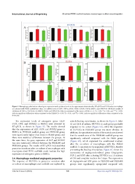
Figure 3. Macrophage polarization-related gene expression and cytokines levels in the supernatants determined by RT-qPCR and ELISA after macrophage
co-cultured with PEEK scaffold for 3 days. (A) mRNA level of CCR7, TNF-α, iNOS, VEGF, CD206, TGF-β, BMP-2, and PDGF-bb. (B) ELISA results of
cytokines of TNF-α, TGF-β, IL-10, BMP-2, VEGF, and PDGF-bb. Data are expressed as the mean ± SD for n ≥ 3; *p < 0.05, **p < 0.01, and ***p < 0.001
indicate significant differences when compared to the PEEK0; p < 0.05, p < 0.01, and p < 0.001 indicate significant differences when compared to the
#
##
###
PEEK200.
The expression levels of osteogenic genes (ALP, scratch healing experiments, as shown in Figure 6. After
OCN, OPN, and RUNX2) in BMSCs were detected by 12 and 24 h of culture, HUVECs in each group gradually
RT-qPCR, as shown in Figure 5E. The results showed migrated to the center (Figure 6A), while the migration
that the expressions of ALP, OCN, and RUNX2 genes in of HUVECs in PEEK400 group was more obvious. In
BMSCs in PEEK400 scaffold group and PEEK200 group addition, the quantitative results of the scratch area showed
were significantly higher than those in PEEK0 group, and that the scratch area of the PEEK400 scaffold group was
there were statistical differences between the groups. At significantly reduced compared with the PEEK group
the same time, the gene expression of ALP and RUNX2 (Figure 6B), which explained the conditioned medium
was also statistically different between the PEEK200 and after the co-culture of macrophages with the PEEK0
PEEK400 groups. The results of RT-qPCR indicated that scaffold. It can promote the migration of HUVECs, thereby
paracrine secretions after co-culture of macrophages with promoting the healing of scratches. Figure 6C shows the
macropore-sized PEEK scaffolds could mediate the high expressions of angiogenesis-related genes (Angiogenin,
expression of osteogenic genes in BMSCs.
FGF-2, and SDF) after culturing HUVECs in a 1:1 mixture
3.4. Macrophage-mediated angiogenic properties of CM and complete medium for 3 days. The expressions
The response of HUVECs to paracrine secretion after of Angiogenin and SDF genes in PEEK200 and PEEK400
co-culture of macrophages and scaffolds was explored by groups were significantly upregulated compared with
Volume 9 Issue 5 (2023) 135 https://doi.org/10.18063/ijb.755