
Page 149 - IJB-9-5
P. 149
International Journal of Bioprinting 3D printed PEEK scaffold mediates macrophages to affect osseointegration
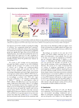
Figure 10. Schematic diagram of 3D-printed PEEK scaffolds with different pore sizes regulating macrophage polarization to promote osseointegration.
Macrophages can respond to the changes of scaffold pore size, and the increase in scaffold pore size can promote macrophage M1 to M2 phenotype transition,
while promoting angiogenesis of HUVECs and osteogenic differentiation of BMSCs in vitro, and subsequently enhancing scaffold osseointegration.
the migration of HUVECs, thereby promoting the healing and stiffness of the PEEK400 scaffold were higher, which
of scratches. The angiogenesis-related gene expression further indicated that the PEEK scaffold with larger pore
results of HUVECs cultured in CM for 3 days showed that size had better osseointegration performance with the host
the expressions of Angiogenin and SDF genes in PEEK400 bone.
group were significantly higher than those in PEEK0 and Macrophages are sensitive to the pore size of the
PEEK200 groups. The results indicated that the CM of the scaffold, and PEEK scaffolds with larger pore size can
scaffold group with larger pore size had a significant in induce the polarization of macrophages from M1 type
vitro pro-angiogenic effect, which was closely related to the to M2 type, and promote the angiogenesis of HUVECs
vascular-associated PDGF-bb growth factor released by and the osteogenic differentiation of BMSCs in vitro
macrophages [12,34,42] .
(Figure 10). Furthermore, the PEEK scaffold with larger
The results of ALP staining and its quantification pore size promotes the formation and growth of new
indicated that BMSCs cultured in the CM of PEEK400 bone, thereby achieving a better osseointegration effect.
scaffolds had the strongest ALP activity, and the trend Therefore, the results of this study demonstrated that
of Alizarin red S staining and osteogenesis-related the strategy of mediating macrophages to promote
gene expression results were consistent with the ALP osseointegration of PEEK scaffolds by regulating the pore
results. Because PEEK400 scaffold group can promote size of the scaffolds is feasible, but future research based on
macrophage polarization and secrete high concentrations bionic design of natural bone tissue and precise control of
of osteogenesis-related cytokines (BMP-2, PDGF-bb, etc.), material structure is warranted. In addition, the reason for
we believe that the immune microenvironment induced the M1 to M2 transition mediated by the aperture of 3D
by macrophages co-cultured with PEEK with larger printing PEEK bracket is still unclear. In future research,
pore size is beneficial to the osteogenic differentiation we also need to explore how the pore size of the scaffold, or
of BMSCs [43-45] . Further, the osseointegration effect of the micro-nano multi-level structure of the scaffold, affects
PEEK scaffolds with different pore sizes was investigated other immune cells in the immune system, and then affects
using a rabbit tibial defect model. Micro-CT 2D images the integration of PEEK scaffold and host bone.
and 3D reconstruction showed that compared with the
PEEK0 group, the PEEK400 group had significantly more 5. Conclusion
new bone around and inside the material, and its bone
mineral density, BV/TV, and Tb.N values also increased PEEK scaffolds with pore sizes of 0, 200, and 400 μm
significantly. Furthermore, with the increase of the pore size were prepared by FDM 3D printing technology, and the
of the scaffold, there is a greater amount of new and more pore size-mediated macrophage polarization and its
mature bone inside the scaffold (Figure 8). In addition, effects on subsequent angiogenesis, bone regeneration,
the biomechanical results showed that the maximum load and osseointegration were studied. Our data suggest that
Volume 9 Issue 5 (2023) 141 https://doi.org/10.18063/ijb.755