
Page 182 - IJB-9-5
P. 182
International Journal of Bioprinting Functional materials of 3D bioprinting for wound healing
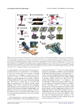
Figure 3. Bioprinting technology. (a) Extrusion bioprinter is a continuous extrusion of cell-containing liquid bioink using manual or pneumatic force.
(b) Schematic diagram of the laser bioprinting device. (c) Schematic illustration of the DLP-based bioprinting device. (d) Inkjet bioprinter sequentially
ejects small droplets of hydrogels and cells to construct tissue. (From ref. [125] licensed under Creative Commons Attribution 4.0 International license.)
(e) Four typical 3D bioprinting techniques correspond to four ways of cutting potatoes. (Reprinted with permission from Gu Z, Fu J, Lin H, et al.,
2020, Development of 3D bioprinting: From printing methods to biomedical applications. Asian J Pharm Sci, 15(5):529–557 [123] . Copyright © 2019
Shenyang Pharmaceutical University.) (f ) Rendered image of the handheld skin bioprinter. (f ) Picture of the 3D-bioprinted microfluidic box. (Reprinted
1
2
with permission from Hakimi N, Cheng R, Leng L, et al., 2018, Handheld skin printer: In situ formation of planar biomaterials and tissues. Lab Chip,
18(10):1440–1451 [126] . Copyright © The Royal Society of Chemistry 2018.)
manipulating light to induce the bioink in the exposed area in the form of droplets [106,117-120] (Figure 3d). This bioprinting
to polymerize and cure a complete layer [115] . As the platform techniques can generally be divided into two types: thermal
is raised and lowered, each new cured layer is bonded inkjet bioprinting and piezoelectric inkjet bioprinting [104,121] .
to the previous one, resulting in a complex and smooth A major advantage of inkjet bioprinting is high resolution
structure [116] (Figure 3c). DLP bioprinting technology has (50 µm), which enables the fabrication of complex scaffolds
high printing speed (printing time of seconds to minutes) by printing multiple materials with high fidelity into
and high resolution (200 nm–6 µm) with shorter printing relevant dimensional structures [116] . In addition, it has
time. Furthermore, it enables the use of bioinks with the advantages of high printing speed (10,000 drops per
high cell concentrations (>10 cells/mL) without causing second), simultaneous printing of multiple ink cartridges,
6
clogging of the nozzles [116] . Because of these advantages, and low technology cost [122] . At the same time, inkjet
this technology can simulate the precise structure and cell bioprinting also has some limitations. For example, its small
viability of natural tissues, leading to breakthroughs in the nozzle diameter and easy clogging limit its ability to print
printing of functional living organ structures. However, bioinks with high cell concentration and high viscosity [116] .
DLP printing can only use photocurable bioinks, and the Additionally, exposure of cells to high temperature of the
UV light used during polymerization may have an impact nozzle and shear stress also reduces cell viability [122] . These
on cell viability. four typical 3D bioprinting processes correspond to the
Inkjet bioprinting is a noncontact printing process in inverse processes of potato slicing, shredding, dicing, and
which bioinks loaded into nozzles are stacked into structures mashing, respectively [123] (Figure 3e).
Volume 9 Issue 5 (2023) 174 https://doi.org/10.18063/ijb.757