
Page 184 - IJB-9-5
P. 184
International Journal of Bioprinting Functional materials of 3D bioprinting for wound healing
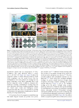
Figure 4. 3D bioprinting of functional wound dressings. (a) Preparation scheme of the Gel/Alg/HA/PPV bioink. (b) Photograph of the 3D bioprinting
process. (c) Flexibility testing of the 3D-bioprinted skin patch. (d) “ICCAS” alphanumeric picture printed with the Gel/Alg/HA/PPV bioink. (e) Pictures
of S. aureus-infected wounds treated with skin patch for 30 min on days 1 and 4 following infection of the rat wound. (f) Photographs of colony forming
units on S. aureus agar plates at infected sites in rats treated with different skin patches. (Reprinted with permission from Zhao H, Xu J, Yuan H, et al., 2022,
3D printing of artificial skin patches with bioactive and optically active polymer materials for anti-infection and augmenting wound repair. Mater Horiz,
9(1):342–349 [130] . Copyright © The Royal Society of Chemistry 2022.) (g) Pictures of agar plates of S. aureus and E. coli O157:H7 colonies treated with
different GCX dressings for different times. (h) SEM images of 3D-printed GCX-CeO /APSGH-Cl dressings with blood cell adhesion. **p ≤ 0.01 (Reprinted
2
with permission from Yang Z, Ren X, Liu Y, et al., 2021, N-halamine modified ceria nanoparticles: Antibacterial response and accelerated wound healing
application via a 3D printed scaffold. Compos Part B-Eng, 227:109390 [131] . Copyright © 2021 Elsevier Ltd). (i) 3D-printed intelligent bandages that undergo
wound scanning, computer modeling and personalization for accurate wound identification and treatment. (j) 3D-bioprinted gelatin hydrogel as an active
ingredient encapsulated on a medical bandage to assemble an intelligent bandage. (k) SEM image of the gelatin hydrogel microstructure. (l) H&E-stained
and (m) MTC-stained sections of wound tissue on day 10 after treatment with different dressings: (i) untreated procedure; (ii) mismatched bandages;
(iii) the intelligent bandages. Scale bar: 100 μm (Reprinted with permission from He X, Yang S, Liu C, et al., 2020, Integrated wound recognition in
bandages for intelligent treatment. Adv Healthcare Mater, 9(22):2000941 [132] . Copyright © 2020 Wiley-VCH GmbH).
experiments showed that the incorporation of CeO / and complete care [132] . Traditional wound dressings often
2
N-halamine NPs could effectively inhibit S. aureus have excessive or incomplete coverage of the wound bed,
and E. coli O157: H7 (Figure 4g). Figure 4h shows the which can lead to suboptimal care outcomes [126] . Therefore,
coagulation ability of various dressings tested by whole precision medicine and personalized treatment are very
blood coagulation test, the results showed that the important [143,144] . Some studies have developed intelligent
3D-bioprinted GCX-CeO /APSGH-Cl dressing had the bandages for precise wound treatment through image
2
lowest coagulation index (BCI), indicating that it had recognition technology, computer modeling, and custom
the best coagulation ability. In addition, compared with material fabrication (Figure 4i and 4j). The intelligent
traditional dressings, 3D-bioprinted dressings had fast bandage prints the gelatin hydrogel using computer
water absorption and better swelling properties. Mouse modeling connected to a bioprinter after quickly obtaining
skin wound repair model and histological analysis the wound geometry via a scanner or smartphone. Scanning
demonstrated that the GXC-CeO /APSGH-Cl dressing electron microscopy revealed 3D-bioprinted hydrogels
2
could promote the regeneration of epidermal and dermal, with typical scaffold microstructures after crosslinking with
thereby promoting wound healing. 3-[cyano(ethyl)amino]propyldimethylazanium chloride/N-
In the process of wound management, wound dressings hydroxysuccinimide (EDC/NHS) (Figure 4k). The addition
that match irregular wounds can provide more precise of silver nanoparticle (AgNP) antimicrobial carrier endowed
Volume 9 Issue 5 (2023) 176 https://doi.org/10.18063/ijb.757