
Page 188 - IJB-9-5
P. 188
International Journal of Bioprinting Functional materials of 3D bioprinting for wound healing
pro-inflammatory chemokines and persistently high levels reports of 3D-bioprinted tissue-engineered scaffolds for
of ROS at the wound site, hindering skin repair [155] . The wound healing.
addition of anti-inflammatory materials to 3D-bioprinted
scaffolds can produce wound healing materials that 6. Discussion and future perspectives
enhance the ability of inflammatory inhibitors, reduce This paper introduces various bioprinting methods,
pro-inflammatory chemokines, eliminate ROS, and functional materials, and their applications in wound
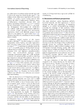
promote macrophage polarization . Li et al. [147] reported dressing and skin tissue engineering. 3D bioprinting
[42]
the anti-inflammatory activity of PDA in a rat model. emerges as an additive bio-manufacturing technique
The 3D-bioprinted PDA-modified BC (DOPA-BC) possessing the advantages of high resolution, flexible
skin scaffolds could prevent inflammatory infiltration operation, repeatable fabrication, and high-throughput
and promote collagen deposition and microvascular output for printing the intricate 3D structures that match
regeneration, which could effectively promote wound the geometric shape of skin wound [117,158] , thus it has been
repair in a rat diabetic skin repair model (Figure 6g). widely used in wound dressings and skin tissue engineering
Therefore, this DOPA-BC scaffold may be ideal for treating scaffolds in recent years [19,43] . As one of the development
diabetic wounds. trends of advanced materials, multifunctional materials
Traditional surgical resection of skin tumors have become an attractive option for wound dressings and
remains a challenge [156] . The ideal strategy is to enhance skin tissue engineering scaffolds. However, the cytotoxicity
postoperative wound healing and tissue regeneration that may occur when the dosage of these multifunctional
while removing residual tumor cells to prevent tumor materials exceeds the cytotoxicity threshold is not
recurrence [32,156,157] . Considering the individual needs, Ma negligible. Moreover, unlike traditional bandages, current
et al. [148] successfully prepared 3D-bioprinted hydrogel 3D-bioprinted hydrogel dressings usually suffer from
scaffolds based on sodium alginate (SA), calcium silicate poor mechanical strength and stability although possess
nanowires (CS), and oligomeric proanthocyanidins multiple functions, and do not function on knees and
(OPC). This CS+SA+4%OPC hydrogel scaffold joints for long periods due to poor adhesion. Also, current
containing photothermal agent OPC could inactivate 3D-bioprinted dressings required more research in
melanoma cells and prevent their growth by controlling overcoming the challenges of scars, nonoxygen permeable
high temperature through NIR irradiation. The in vivo and damaged skin cells [159] .
therapeutic potential of this scaffold was evaluated using The main distinctions of skin tissue engineering
tumor-bearing diabetic mice, and the results are shown compared to the wound dressing are the loaded cells
in Figure 7a–f. Under NIR irradiation, the controllable and bioactive factors. Whether it is to print the bionic
photothermal properties of this scaffold induced high skin structures with cell-encapsulating bioink, or to
temperature to successfully ablate the tumor, so that the inoculate cells on the noncellular-printed scaffolds, the
wound healed without tumor recurrence. In addition, requirements of skin tissue engineering scaffolds for
H&E staining showed epithelialization and collagen printing materials and conditions are stricter than that
deposition (Figure 7g). Therefore, the CS+SA+4%OPC of the printed dressings, such as biocompatibility and
scaffold could effectively treat melanoma and promote viscosity of the bioinks, suitable temperature and pH,
skin wound healing. and sterile microenvironment for cell survival [114,160] .
With the development of 3D bioprinting technology, Although significant progress has been achieved in tissue
flexible polymer materials have been used to build engineering over the years, only a limited number of
complex functional soft structures, that reach a modulus bioinks have the tissue matching characteristics and the
(103–109 Pa) similar to that of human tissues (such as ability to promote tissue generation [161] . At present, it is still
skin or muscle tissue), which is crucial for the process a major challenge for skin tissue engineering to configure
of wound repair . A recent study fabricated porous multifunctional bioink with printability, biocompatibility,
[88]
2,2,6,6-tetramethylpiperidine 1-oxyl (TEMPO)-mediated and excellent mechanical integrity under individual
oxidized cellulose nanofibrils (TCNFs)/casein-based condition [162] . Therefore, the design of mixed bioink should
composite hemostatic scaffolds with cytocompatibility integrate the advantages of natural bioink and synthetic
and hemocompatibility by flexible 3D bioprinting bioink to prepare bioink that is conducive to cell growth
(Figure 7h). Biocompatible TCNF, chitosan and casein and can support cell survival in the printing process [160] . In
with synergistic hemostasis mechanism could endow 3D addition, cell encapsulation bioink can use various types of
composite scaffolds with the ability of cell attachment cells, such as fibroblasts, keratinocytes, mesenchymal stem
and hemostasis (Figure 7i), maximizing their potential cells, and induced pluripotent stem cells, as cell sources [117] .
in wound healing applications . Table 3 summarizes the Stem cells, such as induced pluripotent stem cells, can
[60]
Volume 9 Issue 5 (2023) 180 https://doi.org/10.18063/ijb.757