
Page 190 - IJB-9-5
P. 190
International Journal of Bioprinting Functional materials of 3D bioprinting for wound healing
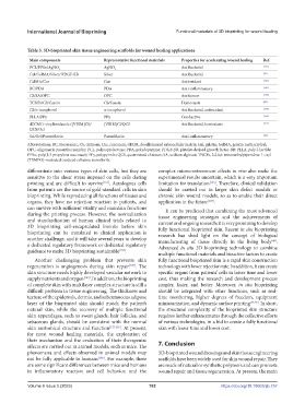
Table 3. 3D-bioprinted skin tissue engineering scaffolds for wound healing applications
Main components Representative functional materials Properties for accelerating wound healing Ref.
PCL/PPSu/AgNO AgNO Antibacterial [145]
3 3
Gel/GelMA/Silver/ PDGF-BB Silver Antibacterial [25]
GelMA/Cur Cur Antioxidant [146]
BC/PDA PDA Anti-inflammatory [147]
CS/SA/OPC OPC Antitumor [148]
TCNFs/CS/Casein CS/Casein Hemostasis [60]
CS/α-tocopherol α-tocopherol Antibacterial, antioxidant [149]
PLLA/PPy PPy Conductive [150]
dECM/1-vinylimidazole ([VBIM]Cl)/ [VBIM]Cl/QCS Antibacterial, hemostasis [151]
QCS/Gel
SA/Gel/Paeoniflorin Paeoniflorin Anti-inflammatory [49]
Abbreviations: BC, bioceramic; CS, chitosan; Cur, curcumin; dECM, decellularized extracellular matrix; Gel, gelatin; GelMA, gelatin methacryloyl;
OPC, oligomeric proanthocyanidin; PCL, polycaprolactone; PDA, polydopamine; PDGF-BB, platelet-derived growth factor-BB; PLLA, poly-l-lactide;
PPSu, poly(1,3-propylene succinate); PPy, polypyrrole; QCS, quaternized chitosan; SA, sodium alginate; TNCFs, 2,2,6,6-tetramethylpiperidine 1-oxyl
(TEMPO)-mediated oxidized cellulose nanofibrils.
differentiate into various types of skin cells, but they are complex microenvironment effects in vivo also make the
sensitive to the shear stress imposed on the cells during experimental results uncertain, which is a very important
printing and are difficult to survive [161] . Autologous cells limitation for translation [166] . Therefore, clinical validation
from patients are the source of gold-standard cells in skin should be carried out in larger skin defect models or
bioprinting. While reproducing all functions of tissues and chronic skin wound models, so as to enable their direct
organs, they have no rejection reaction to patients, and application in the future [167] .
can survive with sufficient vitality and maintain functions It can be predicted that combining the most advanced
during the printing process. However, the normalization tissue engineering strategies and the achievements of
and standardization of human clinical trials related to current and ongoing research; it is very promising to develop
3D bioprinting cell-encapsulated bioinks before skin fully functional bioprinted skin. Recent in situ bioprinting
bioprinting can be translated to clinical application is research has shed light on the concept of biological
another challenge, and it will take several years to develop manufacturing of tissue directly in the living body .
[46]
a dedicated regulatory framework or dedicated regulatory Advanced in situ 3D bioprinting technology to combine
guidance to make 3D bioprinting sustainable [163] .
multiple functional materials and bioactive factors to create
Another challenging problem that prevents skin fully functional bioprinted skin is a rapid skin construction
regeneration is angiogenesis during skin repair [107] . The technology with lower rejection rate. In addition, it can create
skin structure needs highly developed vascular network to specific organs from patients’ cells in lesser time and lower
supply nutrients and oxygen [164] . In addition, the bioprinting cost, thus making the research and development process
of complete skin with multilayer complex structure is still a simpler, faster, and better. Moreover, in situ bioprinting
difficult problem in tissue engineering. The thickness and should be integrated with other functions, such as real-
texture of the epidermis, dermis, and subcutaneous adipose time monitoring, higher degrees of freedom, equipment
layer of the bioprinted skin should match the patient’s miniaturization, and dynamic surface printing [44,46] . In short,
natural skin, while the recovery of multiple functional the structural complexity of the bioprinted skin structure
skin appendages, such as sweat glands, hair follicles, and requires further enhancements through the collective efforts
sebaceous glands, should be consistent with the normal of various technologies, in a bid to create a fully functional
skin anatomical structure and function [117,165] . At present, skin with lesser time and lower cost.
for most wound healing materials, the exploration of
their mechanism and the evaluation of their therapeutic
effects are carried out in animal models, such as mice. The 7. Conclusion
phenomena and effects observed in animal models may 3D-bioprinted wound dressings and skin tissue engineering
not be fully applicable to humans [166] . For example, there scaffolds have been widely used for skin wound repair. They
are some significant differences between mice and humans are made of natural or synthetic polymers and can promote
in inflammatory reaction and cell behavior, and the wound repair and tissue regeneration. At present, the main
Volume 9 Issue 5 (2023) 182 https://doi.org/10.18063/ijb.757