
Page 189 - IJB-9-5
P. 189
International Journal of Bioprinting Functional materials of 3D bioprinting for wound healing
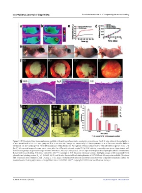
Figure 7. 3D-bioprinted skin tissue engineering scaffolds with antitumor, hemostatic, conductive properties. At 0 and 10 min, infrared thermal pictures
of mice treated with (a) CS+SA+laser group and (b) CS+SA+4%OPC+laser group, respectively. (c) The temperature curve of the tumor site after different
treatments. (d) The volume growth curve of tumor in mice within 14 days. (e) Photographs of tumor of mice treated with different four groups on the 15th
day. (f) Microscopic images of mouse tumor tissue after four different treatments on day 15. (g) H&E staining images of mouse tumor tissue treated with
four different groups. (Reprinted with permission from Ma H, Zhou Q, Chang J, et al., 2019, Grape seed-inspired smart hydrogel scaffolds for melanoma
therapy and wound healing. ACS Nano, 13(4):4302–4311 [148] . Copyright © 2019 American Chemical Society.) (h) Digital images of 3D-bioprinted TCNF
gel model and printing process (h −h ). (i) At 15, 20, 35, and 50 min, the whole blood clotting kinetics of 3D TCNF and 3D composite scaffolds. (Reprinted
2
1
with permission from Biranje SS, Sun J, Cheng L, et al., 2022, Development of cellulose nanofibril/casein-based 3D composite hemostasis scaffold for
potential wound-healing application. ACS Appl Mater Inter, 14(3):3792–3808 . Copyright © 2022 American Chemical Society.)
[60]
Volume 9 Issue 5 (2023) 181 https://doi.org/10.18063/ijb.757