
Page 187 - IJB-9-5
P. 187
International Journal of Bioprinting Functional materials of 3D bioprinting for wound healing
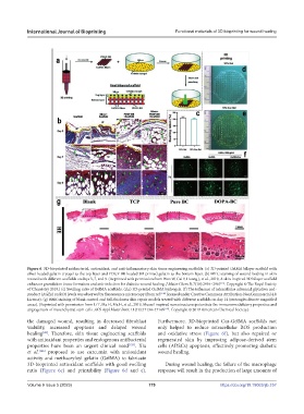
Figure 6. 3D-bioprinted antibacterial, antioxidant, and anti-inflammatory skin tissue engineering scaffolds. (a) 3D-printed GelMA bilayer scaffold with
silver-loaded gelatin cryogel as the top layer and PDGF-BB-loaded 3D-printed gelatin as the bottom layer. (b) MTC staining of wound healing in skin
treated with different scaffolds on days 3, 7, and 9. (Reprinted with permission from Wan W, Cai F, Huang J, et al., 2019, A skin-inspired 3D bilayer scaffold
[25]
enhances granulation tissue formation and anti-infection for diabetic wound healing. J Mater Chem B, 7(18):2954–2961 . Copyright © The Royal Society
of Chemistry 2019.) (c) Swelling ratio of GelMA scaffolds. (d,e) 3D-printed GelMA hydrogels. (f) The influence of intracellular advanced glycation end-
product (AGEs) on ROS levels was observed by fluorescence microscopy (from ref. [146] licensed under Creative Commons Attribution-NonCommercial 4.0
License). (g) H&E staining of blank control and full-thickness skin repair models treated with different scaffolds on day 14 (rectangles denote magnified
areas). (Reprinted with permission from Li T, Ma H, Ma H, et al., 2019, Mussel-inspired nanostructures potentiate the immunomodulatory properties and
angiogenesis of mesenchymal stem cells. ACS Appl Mater Inter, 11(19):17134–17146 [147] . Copyright © 2019 American Chemical Society.)
the damaged wound, resulting in decreased fibroblast Furthermore, 3D-bioprinted Cur-GelMA scaffolds not
viability, increased apoptosis and delayed wound only helped to reduce intracellular ROS production
healing . Therefore, skin tissue engineering scaffolds and oxidative stress (Figure 6f), but also repaired or
[42]
with antioxidant properties and endogenous antibacterial regenerated skin by improving adipose-derived stem
properties have been an urgent clinical need [128] . Xia cells (ADSCs) apoptosis, effectively promoting diabetic
et al. [146] proposed to use curcumin with antioxidant wound healing.
activity and methacryloyl gelatin (GelMA) to fabricate
3D-bioprinted antioxidant scaffolds with good swelling During wound healing, the failure of the macrophage
ratio (Figure 6c) and printability (Figure 6d and e). response will result in the production of large amounts of
Volume 9 Issue 5 (2023) 179 https://doi.org/10.18063/ijb.757