Page 291 - IJB-9-5
P. 291
International Journal of Bioprinting                              Blood components for tissue graft bioprinting


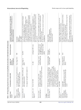
                 Evaluation of the output/biological aspects. Proliferation,


                     rabbit osteochondral lesion. migration and differentiation. In vivo M2 infiltration in a   Proliferation  Migration  Differentiation  Bone rigidity In vitro: Mechanical properties, cell proliferation-osteogenic  differentiation, macrophage polarization. In vivo: Subcutaneous implantation of bone grafts to assess  vascularization, enhanced vascularization with PRP- GA&laponite/PCL; implantation of bone grafts in rat femoral  condyle defects, X-ray and


             Table 1. 3D bioprinting of specific tissue constructs using bioinks functionalized with blood-derived biomaterials


                 Blood-derived product  FFP  Rat PRP  Autologous PRP (4 mL)  PRP, prepared “in house,”   with high platelet count   and high concentration of   VEGF, PDGF-AA, bFGF,   TGF-β3  Fibrinogen  Thrombin (#)  PRP  PRP


                 Modality/cell   phenotype.   Printing/BMSC  Bioprinting/ MSCs  Bioprinting/ rat   BMSC  No cells  3D printing / hASCs   seeded post-printing  Printing bioprinting   / MSCs spheroids  printing/BMSC  Bioprinting/ATDC5   murine chondrocyte   cell line


                 Ink formulation. GelMA/PRP  3 w/v% alginate and 9 w/v% methylcellulose, a  pasty bioink (plasma-Alg-MC) CPC (calcium phosphate cement)  Three bioinks: GA/PCL, PRP-GA/PCL, and   PRP-GA@laponite/PCL GA composite hydrogel: GelMA/AlgMA (5:1)  PCL/β-TCP/PRP  Silk fibroin, HA, gelatin, TCP 3D scaffold printing coated with PRP Sterilization for 2 h using UV and 70% ethanol  PEG 20%–alginate 2.5% hydrogel containing  0.25% photoinitiator and supplemented with  1


                 Author (year);  Journal  Bone  Ahlfeld et al. (2020);  ACS Applied Materials   & Interfaces [47]  Cao et al. (2023); Int J   Bioprint  [48]  Hao et al. (2023) ; Int J   Bioprint  [49]  Wei et al. (2019);   Bioactive Materials  [50]  Cartilage  de Melo et al. (2019);   Adv Funct Mater  [43]  Jiang et al. (2021); Acta   Biomater  [51]  Irmak and   Gümüşderelioglu   (2020); Biomedical   Materials  [52]


            Volume 9 Issue 5 (2023)                        283                         https://doi.org/10.18063/ijb.762

