
Page 474 - IJB-9-5
P. 474
International Journal of Bioprinting 3D printed hydrogel for infected wound healing via PDT
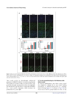
Figure 3. Influence of 3D bioprinted MB@UiO-66(Ce)/PH on the behavior of L929 cells in vitro. (A) The adhesion of L929 cells cultured on the PH-0,
PH-0.1, PH-0.5, and PH-1 hydrogels after 12 h. Scale bar: 100 μm. (B) Quantitative analysis of the proliferation of L929 cells cultured with hydrogels by
CCK-8. Data are means ± SD; n = 3. *p < 0.05, **p < 0.01. (C) In vitro live/dead assay of L929 cells cultured on MB@UiO-66(Ce)/PH, live cells (green), and
dead cells (red). Scale bar: 250 μm.
These results verified the photodynamic antibacterial 3.7. In vivo wound healing in a full-thickness skin
properties of MB@UiO-66(Ce). It is worth noting that the defect model
NPs contain low amounts of MB, as mentioned above. This An infected full-thickness skin defect mouse model
good photodynamic antimicrobial activity also highlights was adopted to evaluate the in vivo wound healing
the fact that utilizing UiO-66(Ce) loaded with MB allows performance of MB@UiO-66(Ce)/PH as a potential
for a more uniform dispersion, which facilitates the wound dressing Figure 6A. As shown in Figure 6B, the
production of O under light. wound areas in all five groups healed with time. At any
1
2
Volume 9 Issue 5 (2023) 466 https://doi.org/10.18063/ijb.773