
Page 476 - IJB-9-5
P. 476
International Journal of Bioprinting 3D printed hydrogel for infected wound healing via PDT
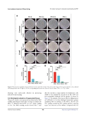
Figure 5. Photodynamics regulation of bacterial behavior in vitro under 660-nm light. Plate colony count images of (A) S. aureus and (B) E. coli co-cultured
with hydrogels under 660-nm light. (C, D) The corresponding antibacterial ratio statistics. Data are means ± SD; n = 4. ****p < 0.0001.
dressings were particularly effective in promoting after the operation, a large number of inflammatory cells
infected wound healing. could be seen around the wound site in the CON and
non-nanoparticle hydrogel (PH-0) groups, as shown in
3.8. Histological evaluation of regenerated tissues Figure 7A. In contrast, there was no obvious inflammatory
The paraffin sections were prepared and observed by H&E cell infiltration in the three hydrogel implantation groups
staining and Masson’s trichrome staining to evaluate the containing NPs. In addition, in the PH-0.1, PH-0.5, and
effect of MB@UiO-66(Ce)/PH on skin defect healing PH-1 groups containing NPs, partial epithelial crawling
in vivo. The H&E staining results showed that 7 days repair was observed on the surface. Compared with the
Volume 9 Issue 5 (2023) 468 https://doi.org/10.18063/ijb.773