
Page 127 - IJB-9-6
P. 127
International Journal of Bioprinting Exosome-based bioink for bioprinting
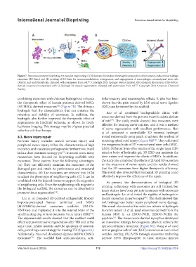
Figure 7. Exosomes promote bioprinting for vascular engineering. (A) Schematic illustration showing the preparation of bioceramic-induced macrophage
exosomes (BC-Exos) and 3D printing of BC-Exos for immunomodulation, osteogenesis, and angiogenesis of macrophages, mesenchymal stem cells
[36]
(MSCs), and endothelial cells. Adapted with permission from ref. . Copyright 2021 Springer Nature Limited. (B) Schematic illustration of hP-MSCs-
[72]
derived exosomes incorporated with CS hydrogel for muscle regeneration. Adapted with permission from ref. . Copyright 2018 American Chemical
Society.
combining exosomes with chitosan hydrogels to enhance inflammatory, and neurotrophic effects. It also has been
the therapeutic effect of human placenta-derived MSCs shown that the pain caused by L5/6 spinal nerve ligation
[72]
(hP-MSCs) derived exosomes (Figure 7B). The chitosan (SNL) can be treated by this scaffold.
hydrogels had the characteristics that can enhance the
retention and stability of exosomes. In addition, the Rao et al. combined biodegradable chitin with
hydrogels also further improved the therapeutic effect of exosomes derived from the gums to treat the sciatic defects
[79]
angiogenesis in hindlimb ischemia, as shown by firefly of rats . The study results showed that exosomes were
luciferase imaging. This strategy may be of great practical effective for treating nerve injuries, and it was a method
value for cell-free therapy. of nerve regeneration with excellent performance. Han
et al. proposed a controllable 3D external hydrogel
4.3. Nerve injury repair mixed microneedle array patch to achieve the method of
[76]
Nervous injury includes central nervous injury and repairing spinal cord injury (Figure 8B) . They cultivated
peripheral nerve injury. It has the characteristics of high the exogenous body of 3D mesenchymal stem cells (MSC-
incidence and inaccurate pathogenesis; furthermore, it still EXO). Different from other studies of the single-layer (2D)
lacks a clear treatment strategy yet . In recent years, many exterior body of hydraulic gel, 3D-EXO can maintain the
[73]
researchers have focused on bioprinting scaffolds with stem nature and improve the effects of MSCs. In addition,
exosomes. These systems have the following advantages: the study also compared the effects of 2D and 3D exosomes
(A) They can effectively maintain the exosomes of the on the treatment of nerve repair, and the results showed
damaged part and retain its performance and structural that the 3D exosomes have higher therapeutic efficiency.
characteristics. (B) The exosomes are released into ECM This result also revealed that biological 3D printing could
to adjust the phenotype of neighboring cells. (C) It can be effectively improve the efficiency of the repair.
combined with the injured tissues to support the migration At present, the demonstrations of biological 3D
of neighboring cells. Once the neighboring cells migrate to printing technology with exosomes are still limited, but
the biological scaffold, the exosomes can be absorbed to many studies have been put into treatment with exosomes
promote tissue regeneration .
[74]
and hydrogels. Liu et al. tested the rigidity of hydrogels in
Liu et al. prepared 3D-printed collagen/silk fibroin/ loaded exosomes in nerve repair . The study showed that
[80]
hypoxia-pretreated human umbilical cord MSCs soft hydrogel can better repair peripheral nerve damage.
(HUCMSCs)-derived exosomes scaffolds (3D-CS- This result also revealed the selection criteria of hydrogels
HMExos) and implanted it into the injured brain of the in nerve repair. Li et al. used exosomes derived from the
small hunting dog to treat traumatic brain injury (TBI) . human MSC in an EXOO-PGEL (EXOO-PGEL) for
[75]
The experimental results showed that the method could peptides . The tissue nerve-derived injury has developed
[77]
effectively promote nerve regeneration after TBI and, at the an innovative strategy for exogenous physical delivery for
same time, inhibit neuritis and the apoptosis of neurotic spinal cord injury treatment (Figure 8C). Wang et al. used
cells, providing a new strategy for treating TBI (Figure 8A). retinal ganglion cells of rats (RGC) exosomes as nano-sized
Additionally, Hsu et al. developed alginate and HUCMSCs vesicles, loading PACAP38 through exosomes anchoring
exosomes . The scaffold had anti-inoculation, anti- peptide CP05 (Exopacap38) to treat external injuries
[78]
Volume 9 Issue 6 (2023) 119 https://doi.org/10.36922/ijb.0114