
Page 124 - IJB-9-6
P. 124
International Journal of Bioprinting Exosome-based bioink for bioprinting
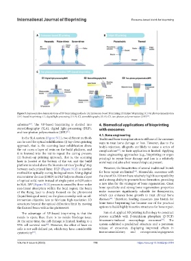
Figure 5. Representative demonstrations of 3D bioprinting methods. (A) Extrusion-based 3D printing; (B) Inkjet 3D printing; (C) Vat photopolymerization
(VP)-based bioprinting: C1, digital light processing (DLP); C2, stereolithography (SLA); C3, two-photon polymerization (2PP) .
[61]
[60]
substrate . The VP-based bioprinting is divided into 4. Biomedical applications of bioprinting
stereolithography (SLA), digital light processing (DLP), with exosomes
[61]
and two-photon polymerization (2PP) .
4.1. Bone engineering
In the SLA system (Figure 5C1), two different methods Traditional bone transplantation is still one of the common
can be used for optical solidification: (i) top-down printing ways to treat bone damage or loss. However, due to the
approach, that is, the scanning laser solidification above body’s rejection, allografts are likely to cause a series of
the vat cures a layer of resin on the build platform, and complications , so their application is limited. Applying
[64]
it is lowered into the vat to repeat the curing process; tissue engineering approaches (e.g., bioprinting or organ
(ii) bottom-up printing approach, that is, the scanning printing) to repair bone damage and loss is a relatively
laser is located at the bottom of the vat, and the build novel way and also a hot research topic at present.
platform is raised above the bioresin vat via a “peeling” step
between each printed layer. DLP (Figure 5C2) is another However, the bioactivities of several traditional bioink
[65]
method for optically curing biological resin. Using digital for bone repair are limited . Meanwhile, exosomes with
micromirror devices (DMD) in DLP helps to obtain a layer the size of 50–120 nm have relatively high biocompatibility
of optical solid resin instead of single-point solidification and a strong ability to promote bone formation, providing
in SLA. 2PP (Figure 5C3) process is caused by three-order a new idea for the strategies of bone regeneration. Great
non-linear absorption within the focal region; the beam bone specificity and strong bone regeneration properties
of the flying laser is closely focused on the photoresist make exosomes significantly valuable for therapeutics,
(liquid biological resin) on the glass coverslip with an oil- which can enhance bone growth to treat clinical bone
[66]
immersion objective lens to fabricate high-resolution 3D diseases . Therefore, loading exosomes into bioink for
structures beyond the optical diffraction limit by moving bone tissue bioprinting has become one of the practical
the focused beam within the photoresist. options to build highly bioactive structures for bone repair.
The advantage of VP-based bioprinting is that the Sun et al. applied 3D printing technology to construct
nozzle is open; thus, there is no nozzle blockage issue. porous scaffolds with β-tricalcium phosphate (β-TCP)
[36]
At the same time, the cell damage is limited, leading to a bioceramic-induced macrophage exosomes . The
95% cell survival rate . However, the effect of laser on system exhibited a predefined structure and a persistent
[62]
cells is not well-defined yet, which may have considerable release of exosomes, displaying improved effects in
cytotoxicity . immunomodulatory and osteogenesis/angiogenesis
[63]
Volume 9 Issue 6 (2023) 116 https://doi.org/10.36922/ijb.0114