
Page 215 - IJB-9-6
P. 215
International Journal of Bioprinting Multi-Cellular tissues/organoids manufacturing strategies
emerged . The advantages of the scaffold-free strategy
[53]
compensate for the limitations of scaffold-based strategies.
The scaffold-free strategy is particularly suitable for
fabricating complex heterogeneous structures containing
multiple cell types [54,55] . This bottom-up assembly approach
holds promise as a module for constructing intricate Bioprinting-assisted tissue emergence Enables spontaneous formation of Precise control over initial spatial organization and density of building Does not require overly complex Placement of spheres in the scaffold Requires suitable conditions and
MTOs . The key bioassembly technologies based on the post-printing remodeling for
[56]
scaffold-free strategy include the Kenzan and aspiration- bioprinting modalities is done manually geometric complexity
assisted technology, fluid-based manipulation, suspended larger MTOs
bioprinting, and bioprinting-assisted tissue emergence. 100–500 µm 10–50 μm/s units
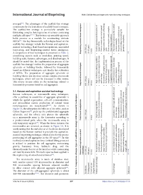
A comparison of four techniques is presented in Table 2, 12 mm • • • • • [129]
considering aspects such as resolution, printing speed,
building scale, features, advantages, and disadvantages. It
should be noted that the implementation process of the
scaffold-free strategy involves the preparation of aggregate
spheroids or building blocks, followed by bioassembly
based on different techniques, and, finally, the cultivation High-resolution printing capability Long-term culture support for bone Generation of complex architectures Perfusion of living matrix with Compatibility with various organoids Limited biomimicry due to dense
of MTOs. The preparation of aggregate spheroids or Suspended-based techniques Challenges in achieving full
building blocks also involves various complex microscale oxygenated media nature of the bioinks endothelialization
techniques, which will not be discussed in this review. and cartilage
This review focuses solely on the technology related to 100–180 µm 5.6 mm/s
bioassembly processes based on cell aggregates. 40 mm • • • • • • • [126–128]
3.1. Kenzan and aspiration-assisted technology
Kenzan techniques, or microneedle array techniques,
offer a pathway to assemblies of aggregate spheroids in
which the spatial organization, cell–cell communication,
and extracellular matrix production of natural tissue Limitations with large cell numbers and Size constraints in optically induced Technical challenges for each technique
morphogenesis are recapitulated [43,57] . As shown in Essential for structural fabrication dielectrophoresis (ODEP) devices
Figure 5A, the adsorption end effector of the robot, such as High biocompatibility Biocompatibility concerns
Cyfuse Regenova , picks up the prefabricated aggregate Fluid-based manipulation Microscale accuracy thicker tissues
[58]
spheroids, and the robotic arm pierces the spheroids 1 nm–300 µm
on a microneedle array in the bioreactor according to 10 mm
a predetermined path, where the microneedle array is - • • • • • • • [125]
only temporary support . When the tissue matures, the
[35]
microneedles are removed, as shown in Figure 5A. It is
worth noting that the end effector of the device developed
based on the Kenzan method is precisely the aspiration-
assisted bioprinting technique, which allows for the precise Table 2. Comparison of major types of scaffold-free strategy with bioassembly techniques
positioning of cell aggregates in the 3D space . In this Precise positioning of cell aggregates Potential for organ pre-vascularization Lower resolution for unknown cell combinations and culture conditions Manual positioning inefficiency at the
[59]
method, minimal aspiration force (critical lifting pressure) Recapitulation of natural tissue Large-scale structure generation Physiological alterations due to spheroid size and compaction
is utilized to position the cell aggregates, overcoming Kenzan and aspiration-assisted
gravity, buoyancy force, hydraulic drag, and the morphogenesis single-cell scale
thermodynamic barrier at the interface while maintaining technology 10–200 µm
cell viability above 80%. This technique has been applied in 400 µm/s 12 mm [43,105,124]
the engineering of osteochondral tissues . • • • • • • •
[59]
The microneedle array is made of stainless steel,
with needles around 150 microneedles in diameter and
500 microneedles spacing between adjacent needles (longest dimension) Feature and advantage
to allow contact with adjacent aggregate spheroids . Printing speed Construct size Disadvantage
[60]
The diameter of the cell-aggregated spheroids is about Resolution Reference
140–500 microneedles [57,61] . The diameter and geometric
Volume 9 Issue 6 (2023) 207 https://doi.org/10.36922/ijb.0135