
Page 211 - IJB-9-6
P. 211
International Journal of Bioprinting Multi-Cellular tissues/organoids manufacturing strategies
section, we compared the advantages, disadvantages, and
applicability of scaffold-based and scaffold-free strategies.
We believe that a balanced consideration of both approaches
is necessary to achieve the common goal of manufacturing
functional tissue-like structures (MTOs). Subsequently,
we delved into the discussion of convergence strategies
and hybrid biofabrication technologies. The vision for
future prospective hybrid technology involves emerging
convergence strategies for fabricating complex MTOs.
Subsequently, a novel MTOs biofabrication method based High resolution (lateral around 20–50 μm, vertical around 25–100 μm) Faster printing speed and the ability to print large-scale structures with
on the convergence strategy, the BioMicroMesh method Ability to print a wide range of viscosities of bioresin (up to 5 Pa·s) Potential to fabricate highly complex structures, support structure Harmful effects of UV rays, resulting in more DNA damage Cytotoxic effects from increased light intensity and photoinitiator
(BMMm), is introduced for the first time. Concurrently, Material degradation and bubble damage in two-photon
the development of new MTOs biofabrication equipment,
the BioMicroMesh system (BMMs), is underway, aiming to
provide precise experimental variable control and ensure polymerization (2PP) technology
the fabrication of commercial MTOs models for biological Vat photopolymerization-based Medium (200–1600 mm/s) Chemical, photocrosslinking fabrication possible micron-level resolution
research. It is hoped that this review will encourage further concentration
research and development in MTOs manufacturing and 10–100 µm >10 8 cells/ml
related specialized equipment. No >95% • • • • • • • [121,123]
2. Scaffold-based strategy with
3D bioprinting techniques
Scaffold-based strategy is a pivotal approach in the
fabrication of MTOs constructs, as scaffolds form
the fundamental architecture of such constructs, and
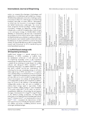
3D bioprinting technology serves as the mainstream Wide range of printable biocompatible materials High mechanical pressure and shear stress
methodology for scaffold manufacturing [19,20] . Scaffolds play Chemical, photocrosslinking, shear dilution, Printable high-viscosity biomaterials Highly controlled printing structure
a crucial role in providing structural support for cellular leading to reduced cell viability
adhesion and subsequent MTOs maturation [21,22] . 3D Most commonly used Limited resolution range
bioprinting technology is an additive fabrication method Extrusion-based Slow (10–50 μm/s)
that prints scaffolds layer-by-layer automatically. The 100–500 µm 10 8 –10 9 cells/ml temperature
technology has become a promising strategy in cell-laden Exist 80% • • • • • • [121,122]
tissue engineering or MTOs restoration [12,23] . In a general
sense, 3D bioprinting can be divided into four development
stages . Stage I involves the printing of non-biocompatible
[24]
structures that can be used as models for surgery planning.
Stage II encompasses the printing of biocompatible but
non-biodegradable products, such as implanted prostheses.
Stage III focuses on the printing of biocompatible and Requires the printed biomaterial to be in liquid Cannot print high-viscosity materials or high
biodegradable products that can serve as scaffolds to Table 1. Comparison of major types of scaffold-based 3D bioprinting techniques Can be equipped with multiple nozzles The thermal actuator is potentially prone to reduced equipment structural stability and Printhead is prone to wear and clogging.
enhance tissue damage repair or regeneration. Finally,
Stage IV involves cellular printing to create biomimetic Chemical, photocrosslinking Complex nozzle structure compromised cell viability. concentrations of cells
3D structures using cells . In this article, we provide a Easy accessibility Low drive pressure
[24]
comprehensive definition of bioprinting that encompasses Jetting-based Fast (1–10 4 drop/s) 10 6 –10 7 cells/ml
the stages of development from Stage III to Stage IV. Based Exist 50–100 µm >85% form [118–120]
on the different printing mechanisms, 3D bioprinting • • • • • • • •
technology can be categorized into three main types: jetting-
based , extrusion-based , and vat photopolymerization-
[25]
[20]
based . Table 1 summarizes the characteristics of three Crosslinking method
[26]
techniques in terms of printhead, printing speed, resolution, Printing speed Cell viability
cell viability, cell density, crosslinking method, features, Nozzle Resolution Cell density Features Limitations Reference
advantages, and disadvantages.
Volume 9 Issue 6 (2023) 203 https://doi.org/10.36922/ijb.0135