
Page 249 - IJB-9-6
P. 249
International Journal of Bioprinting 3D-Printed GelMA biomaterials in cartilage repair
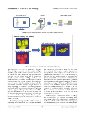
Figure 2. Process of constructing a scaffold with three layers using 3D printing technology.
Figure 3. Construction of 3D computer model for articular cartilage defects.
wave-like interface structure of the calcified cartilage layer and components, especially the method of extrusion-
allows for tight connection with the hyaline cartilage, based printing that can print complex, high-precision
while the rough, serrated interface structure anchors it to biological tissue, which is a good candidate to be used for
the subchondral bone. This unique interface connection cartilage tissue engineering [4,7,8] . With computer assistance,
increases both the contact area and the connection the structures and components can be customized and
strength between articular cartilage interfaces. The printed easily with the aid of different nozzles (Figure 2).
subchondral bone is anchored to each other; this special Meanwhile, the construction of the interface structure of
interface connection method increases the connection complex tissues also utilizes the advantages of 3D printing
area between the articular cartilage interface and also technology. A computer 3D joint model with complex
increases the connection strength. In light of this, it is structure can be constructed (Figure 3), and then different
almost impossible to reconstruct this structure through layers can be printed without extra bonding or splicing
traditional scaffold fabrication techniques, but 3D printing required in traditional scaffold fabrication techniques.
technique appears to be a possible and easy method for Thus, compared to traditional scaffold fabrication
reconstructing this structure. In addition, reconstructing technique, 3D printing offers a rather easy method to
the structures and mimicking the components in different reconstruct this structure.
layers of the articular cartilage, which is a big challenge for Anatomically, the different layers of articular cartilage
traditional scaffold fabrication techniques, can ensure the are heterogeneous. 3D printing technology, rapid
function recovery. prototyping, and an effective personalized manufacturing
3D printing technology provides a viable solution to method of scaffolds can be constructed by superimposing
fabricating multi-layer tissues with complex geometries customized biological inks according to different patients.
Volume 9 Issue 6 (2023) 241 https://doi.org/10.36922/ijb.0116