
Page 286 - IJB-9-6
P. 286
International Journal of Bioprinting Progress in bioprinted ear reconstruction
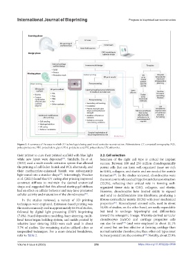
Figure 3. A summary of the ways in which 3D technology is being used to aid auricular reconstruction. Abbreviations: CT, computed tomography; PCL,
polycaprolactone; PEG, polyethylene glycol; PLA, polylactic acid; PU, polyurethane; UV, ultraviolet.
their printer to cure their printed scaffold with blue light 3.2. Cell selection
while new layers were deposited . Similarly, Jia et al. Selection of the right cell type is critical for implant
[14]
(2022) used a multi-nozzle extrusion system that allowed success. Between 100 and 250 million chondrogenically
the printing of cell-laden bioink and PCL alternately, and potent cells that can form well-organized tissue are rich
their methacrylate-enhanced bioink was subsequently in GAG, collagens, and elastin and are needed for auricle
light-cured into a sturdier shape . Interestingly, Visscher formation . In the studies reviewed, chondrocytes were
[11]
[29]
et al. (2021) found that UV curing after printing improved the most commonly used cell type for auricle reconstruction
construct stiffness to maintain the desired anatomical (33.3%), reflecting their critical role in forming well-
shape and suggested that this altered starting gel stiffness organized tissue rich in GAG, collagens, and elastin.
had an effect on cellular behavior and may have promoted However, chondrocytes have limited ability to expand
cellular activity and maturation of the chondrocytes . and tend to dedifferentiate into fibroblasts, producing a
[28]
In the studies reviewed, a variety of 3D printing fibrous extracellular matrix (ECM) with poor mechanical
[23]
techniques were employed. Extrusion-based printing was properties . Mesenchymal stromal cells, used in about
the most commonly used in approximately 18.5% of studies, 14.8% of studies, on the other hand, are easily expandable
followed by digital light processing (DLP) bioprinting but tend to undergo hypertrophy and differentiate
(7.4%). Fused deposition modeling, laser sintering, multi- toward the osteogenic lineage. Microtia-derived auricular
head tissue/organ building system, and molds printed by chondrocytes (mACs) and cartilage progenitor cells
[29]
selective laser sintering (SLS) were each used in about can also be used (and were employed in about 11.1%
3.7% of studies. The remaining studies utilized other or of cases) but are less effective at forming cartilage than
unspecified techniques. For a more detailed breakdown, normal auricular chondrocytes; thus, other cell types must
refer to Table 2. be incorporated into the construct . Notably, about 25.9%
[23]
Volume 9 Issue 6 (2023) 278 https://doi.org/10.36922/ijb.0898