Page 288 - IJB-9-6
P. 288

International Journal of Bioprinting                                Progress in bioprinted ear reconstruction


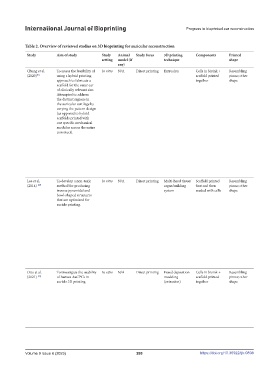
            Table 2. Overview of reviewed studies on 3D bioprinting for auricular reconstruction
             Study       Aim of study        Study   Animal   Study focus  3D printing   Components  Printed                     Printed   Cell nature/type  Notable post-  Assessment   Findings                    Limitations and suggested
                                             setting  model (if        technique                   shape                         material                printing       of success/                                  improvements
                                                   any)                                                                                                  modifications  integration
             Chung et al.   To assess the feasibility of   In vitro  N/A  Direct printing  Extrusion  Cells in bioink +   Resembling   PCL  GelMA-HAMA   Photocuring of the   Histopathology  •  The design of a print affects its stiffness and   •  Further improvements
             (2020) [9]  using a hybrid printing                                     scaffold printed   pinna; other                       cell-supportive   printed bioink was       flexibility significantly.       could be developed
                         approach to fabricate a                                     together      shape                                   bioink        using a 400 nm UV          •  Scaffolds printed with a 400 μm nozzle tip   to generate smoother
                         scaffold for the outer ear                                                                                                      source at a focal            had the lowest compressive modulus but were   intersections for each
                         of clinically relevant size.                                                                                                    distance of 5.0 cm           similar in stiffness to native auricular cartilage   region
                         Attempted to address                                                                                                            and an intensity of          and had the fastest printing time.  •  Incorporate a gradual
                         the distinct regions in                                                                                                         15–30%                     •  Increasing the nozzle diameter decreases   decrease or increase of
                         the auricular cartilage by                                                                                                                                   the compressive modulus of PCL scaffolds,   the strand spacing of
                         varying the pattern design                                                                                                                                   while increasing strand spacing and using   each part at the junction
                         (as opposed to hybrid                                                                                                                                        orientations of 0/45° leads to more flexible
                         scaffolds printed with                                                                                                                                       structures.
                         one specific mechanical                                                                                                                                    •  Scaffolds can serve as a temporary support
                         modulus across the entire                                                                                                                                    for host tissue integration and chondrocyte
                         construct).                                                                                                                                                  differentiation. A scaffold with similar
                                                                                                                                                                                      properties before implantation could improve
                                                                                                                                                                                      its handling and shape retention after
                                                                                                                                                                                      implantation.
                                                                                                                                                                                    •  Cells remained viable for up to 7 days after
                                                                                                                                                                                      printing.
                                                                                                                                                                                    •  The presence of a surrounding hydrogel
                                                                                                                                                                                      during the printing process helps protect cells
                                                                                                                                                                                      from shear forces at the nozzle tip, leading to
                                                                                                                                                                                      good cell viability during the printing process.
             Lee et al.    To develop a non-toxic   In vitro  N/A  Direct printing  Multi-head tissue/  Scaffold printed   Resembling   PCL & PEG  Human ASCs   The cell-printed   Mechanical   •  The sacrificial layer technique allowed for the   A system for incubating
             (2014)  [27]  method for producing                        organ building   first and then   pinna; other                      exposed to    structures were   testing; electron   construction of complex structures of any   printed cells will need to
                         inverse pyramidal and                         system        seeded with cells  shape                              chondrogenic   incubated a   microscopy    shape.                         be developed and attached
                         bowl-shaped structures                                                                                            induction medium  week at 37℃            •  The PEG sacrificial component did not   to the printer in order to
                         that are optimized for                                                                                            and adipogenic   in a humidified           impact cell viability or proliferation and could  keep the cells alive during
                         auricle printing.                                                                                                 induction medium  atmosphere               be easily dissolved in water or cell culture   the printing of large
                                                                                                                                                         containing 5% CO .           media within 40 minutes.       structures such as an ear, as
                                                                                                                                                                     2
                                                                                                                                                         The sacrificial PEG        •  Hydrogels may provide a better environment   the viability of the printed
                                                                                                                                                         layer was dissolved.         for chondrocyte proliferation, but printed   cells may be compromised
                                                                                                                                                                                      adipocytes had a lower proliferation rate.  during this process.
                                                                                                                                                                                    •  Chondrocytes and adipocytes had similar
                                                                                                                                                                                      proliferation rates when printed separately,
                                                                                                                                                                                      and chondrogenesis and adipogenesis
                                                                                                                                                                                      occurred effectively when the two cell types
                                                                                                                                                                                      were co-printed and co-cultured.
                                                                                                                                                                                    •  An ear-shaped structure containing both
                                                                                                                                                                                      chondrocytes and adipocytes not only
                                                                                                                                                                                      maintained its shape, but also regenerated
                                                                                                                                                                                      both auricular cartilage and earlobe fat.
             Otto et al.    To investigate the usability   In vitro  N/A  Direct printing  Fused deposition   Cells in bioink +   Resembling   PCL  Novel human   Cultured in vitro   Histopathology;  •  Extrusion printing does not negatively   Short-term study; no in vivo
             (2021)  [29]  of human AuCPCs in                          modeling      scaffold printed   pinna; other                       AuCPCs        in chondrogenic   micro-CT scan;   impact cell viability, metabolic activity, or the   testing of this method yet
                         auricle 3D printing.                          (extrusion)   together      shape                                                 media for 30 days  mechanical   production of GAGs.
                                                                                                                                                                        testing     •  The extrusion of AuCPCs through a
                                                                                                                                                                                      microvalve system did not harm cell viability
                                                                                                                                                                                      (which remained at 8% over 10 days),
                                                                                                                                                                                      metabolic activity (which was not different
                                                                                                                                                                                      between cast and printed cells), or GAG
                                                                                                                                                                                      production (which occurred over 28 days in
                                                                                                                                                                                      vitro).
                                                                                                                                                                                    •  PCL scaffolds with various strand spacings
                                                                                                                                                                                      support GAG production.


            Volume 9 Issue 6 (2023)                        280                        https://doi.org/10.36922/ijb.0898
   283   284   285   286   287   288   289   290   291   292   293