
Page 282 - IJB-9-6
P. 282
International Journal of Bioprinting Progress in bioprinted ear reconstruction
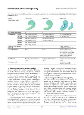
Table 1. An overview of chief differences between landmark surgical methods in auricular autografting (adapted from Dr. Nagata’s
original sketches )
[13]
Method Tanzer (1959) Brent (1980) Nagata (1992)
Costal cartilage frame
Total number of operations 6 4 2
Operation time 1st stage 1 h—Lobule transposition 3 h—Lobule transposition 8 h—Costal cartilage graft
2nd stage 3 h—Costal cartilage graft 1 h—Costal cartilage graft 8 h—Ear projection
3rd stage 2 h—Tagus construction 2 h—Tagus construction
4th stage 2 h—Separating the ear 2 h—Separating the ear
5th stage 1 h—Temporary tunnel
6th stage 2 h—Closing the tunnel
Number of costal cartilages 3 3 4 for 1st stage (ribs 6–9)
harvested 2 for 2nd stage (ribs 4 and 5)
Chest wall deformity Occur Occur Do not occur because the
perichondrium is left in place
and refilled using diced cartilage
remnants from cutting before
closing. Thus, the cartilage
regenerates over time.
Resorption risk High due to insufficient blood High due to insufficient blood No resorption due to good blood
supply supply supply
Wire sutures used 5 5 85 needed for 1st stage
20 needed for 2nd stage
1.1. Current reconstructive surgeon’s toolbox particularly valuable in patients with inadequate available
Current options for treating auricular deformities loco-regional skin, such as in burns or in cases where
or absence include cartilage autografting, alloplastic autologous reconstruction was unsuccessful. They are
polyethylene implants such as Medpor, and prostheses [3,7] . generally a last resort or reserved for patients who prefer
minimally invasive treatment .
[5]
Most (91.3%) surgeons prefer autografting over
alloplasty , partly because autografted cartilage has 1.2. Current surgical techniques in autografting
[7]
a lower risk of being extruded through the skin or of The main reconstructive techniques in sculpting rib
soft-tissue necrosis, whereas Medpor is susceptible to cartilage into an autografted auricle are those by Brent,
minor trauma and secondary infections, dehiscence, and Tazner, and Nagata, with notable variations like that
implant extrusion . Medpor implants typically require by Firmin to improve and adjust results [3,12] . While this
[8]
coverage with a temporoparietal fascial flap to prevent complex reconstruction requires specific surgical and
such complications . Even then, results are suboptimal, artistic skills—thus limiting the field to a small number
[3]
as manufacturing limitations mean implants are not of experienced surgeons—the final result is still often less
customized, and aesthetic results are a compromise at than perfect . This is because the reconstruction is far
[3]
best . Furthermore, the material is inherently stiff and from simple, regardless of technique.
[9]
[10]
easily triggers immune reactions .
[11]
The operations require substantial rib cartilage resection
The alternative route is that of prosthetics, which have (see Table 1). Thus, the initiation of ear reconstruction
evolved considerably in recent years in terms of cosmetic must often be delayed until the child is of an adequate
results. Prostheses can be divided into those applied with a age to have enough usable cartilage. Furthermore, the
silicon adhesive and the surgically osseointegrated. This is procedure involving the chest wall as a donor site can
Volume 9 Issue 6 (2023) 274 https://doi.org/10.36922/ijb.0898