Page 304 - IJB-9-6
P. 304

International Journal of Bioprinting                                Progress in bioprinted ear reconstruction
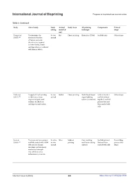


            Table 2. Continued
             Study       Aim of study        Study   Animal   Study focus  3D printing   Components  Printed                     Printed   Cell nature/type  Notable post-  Assessment   Findings                    Limitations and suggested
                                             setting  model (if        technique                   shape                         material                printing       of success/                                  improvements
                                                   any)                                                                                                  modifications  integration
             Dong et al.   To determine the   In vivo   Rat  Direct printing  Extrusion (FDM)  Scaffold only  Other shape        PLA       Bone marrow   Cells were cultured,  Histopathology;  •  A combination of MSCs and chondrocytes   The implantation of the
             (2022)  [48]  minimum fraction   animal                                                                                       ‐derived      passaged, and then   micro-CT scan  was used to create cartilage that demonstrated  constructs in nude rats,
                         of human auricular                                                                                                mesenchymal stem  slowly injected into     consistent mechanical function, even when   which manifest different
                         chondrocyte required                                                                                              cells         the scaffolds and            the ratio of MSCs to chondrocytes was high.  skin characteristics from
                         to form healthy elastic                                                                                                         gelled at 37°C for 1       •  When chondrocytes made up only 10% of   humans (e.g. looseness
                         cartilage when co‐cultured                                                                                                      h. Cell‐loaded               the initial cell population, the resulting tissue   of rodent skin does
                         with human MSCs.                                                                                                                constructs were              had characteristics similar to native elastic   not simulate the high
                                                                                                                                                         then cultured                cartilage in terms of both biomechanics and   pressure caused by
                                                                                                                                                         overnight                    biochemistry.                  implantation under the
                                                                                                                                                         before                     •  Co-implantation of a small number of   tight scalp), as well as
                                                                                                                                                         implantation.                chondrocytes with MSCs in a type I collagen   significant differences in
                                                                                                                                                                                      matrix resulted in the production of cartilage   immunocompetency.
                                                                                                                                                                                      that was indistinguishable from native
                                                                                                                                                                                      auricular cartilage after 6 months in vivo.
                                                                                                                                                                                    •  It is not yet clear if the more efficient cartilage
                                                                                                                                                                                      formation observed in this study is due
                                                                                                                                                                                      to the differentiation of MSCs toward a
                                                                                                                                                                                      chondrogenic lineage, a trophic effect of the
                                                                                                                                                                                      MSCs, or a combination of both, but this is
                                                                                                                                                                                      the subject of ongoing research.
                                                                                                                                                                                    •  The use of a small number of chondrocytes
                                                                                                                                                                                      could be an important step toward the clinical
                                                                                                                                                                                      translation of auricular tissue engineering due
                                                                                                                                                                                      to the limited availability of auricular cartilage
                                                                                                                                                                                      donor tissue.
             Park et al.    To apply 3D cell printing   In vivo   Rabbit  Direct printing  Multi-head tissue/  Cells in bioink +   Other shape  PCL  Primary   Incubated at 37℃   Histopathology;  •  A multi-head tissue/organ building system   Small sample
             (2017)  [49]  to fabricate a tissue-  animal              organ building   scaffold printed                                   chondrocytes from  for 1 h. Fabricated   mechanical   can successfully be used to 3D-print a
                         engineered graft, and                         system (extrusion)  together; scaffold                              the New Zealand   CSHS was   testing; electron   cell-printed structure (CPS) using layers of
                         evaluate its effects on                                     printed first and                                     white rabbit  crosslinked using   microscopy  alginate bio-ink encapsulating chondrocytes
                         cartilage reconstruction.                                   then seeded with                                                    CaCl . Washed                and PCL.
                                                                                                                                                             2
                                                                                     cells                                                               with PBS thrice.           •  The CPS was found to have a higher efficiency
                                                                                                                                                         Then chondrocytes            of cellular settlement, improved survival and
                                                                                                                                                         were seeded onto             function of chondrocytes in vitro compared to
                                                                                                                                                         the CSHS with the            a cell-seeded scaffold (CSS).
                                                                                                                                                         same cell density          •  When implanted in a rabbit ear with a
                                                                                                                                                         with CSS.                    cartilage defect, the CPS led to complete
                                                                                                                                                                                      cartilage regeneration after 3 months, while
                                                                                                                                                                                      the CSS and autologous cartilage only led to
                                                                                                                                                                                      incomplete healing.
                                                                                                                                                                                    •  These results suggest that 3D printing
                                                                                                                                                                                      synthetic polymer scaffolds with hydrogel
                                                                                                                                                                                      materials and cells can be a viable alternative
                                                                                                                                                                                      to using autologous cartilage for auricular
                                                                                                                                                                                      reconstruction.
             Jia et al.    To create and test a proper   In vitro;  Mice  Indirect   Cast-molding   Scaffold printed   Resembling   Other:   Goat chondrocyte   A cell suspension   Histopathology;  •  The scaffold showed excellent   Important unknowns
             (2020)  [25]  scaffold created with ACM   in vivo   printing  and freeze-drying   first and then   pinna; other     ACM/      seeding       was seeded into   electron   biocompatibility and successfully regenerated   remain, including how
                         with precise human-  animal                   mixture       seeded with cells  shape                    gelatin-PCL             each scaffold. This   microscopy  human-ear-shaped cartilage that retained a   best to optimize scaffold
                         ear-shape and necessary                                                                                 scaffold                was followed by 24           satisfactory shape, good elasticity, abundant   preparation, evaluation
                         mechanical strength                                                                                                             h of incubation              lacuna structure, and cartilage-specific ECM   of scaffold biosafety, and
                         that will elicit a low                                                                                                                                       deposition.                    the feasibility of human-
                         inflammatory reaction.                                                                                                                                     •  Cell seeding efficiency in both ACM/gelatin   ear-shaped cartilage
                                                                                                                                                                                      and gelatin scaffolds was more than 90%,   regeneration in large
                                                                                                                                                                                      which was significantly superior than that of   animals.
                                                                                                                                                                                      PGA/PLA scaffolds.


            Volume 9 Issue 6 (2023)                        296                        https://doi.org/10.36922/ijb.0898
   299   300   301   302   303   304   305   306   307   308   309