
Page 299 - IJB-9-6
P. 299
International Journal of Bioprinting Progress in bioprinted ear reconstruction
Study Aim of study Study Animal Study focus 3D printing Components Printed Printed Cell nature/type Notable post- Assessment Findings Limitations and suggested
setting model (if technique shape material printing of success/ improvements
any) modifications integration
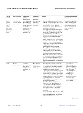
Xia et al. To establish novel scaffold- In vitro; Mice + Direct printing Pneumatic Scaffold printed Resembling Photo- Goat auricular After 3D printing, Histopathology; • Photo-crosslinkable gelatin and HA can be Mechanical strength of the
(2018) [14] fabricated strategies for in vivo goats extrusion-based first and then pinna; other curable cartilage-derived the scaffolds were mechanical fabricated as a porous scaffold with a precise scaffolds warrants further
native polymers and animal bioprinter with a seeded with cells shape hydrogel chondrocytes frozen at −80°C for testing; electron outer shape, good internal pore structure, enhancement, and the
provide a novel natural 3D 405 nm blue light. (meth- 4 h and lyophilized microscopy high mechanical strength, and good feasibility of regenerating
scaffold with satisfactory acrylic for 48 h. The degradation rate, through photocuring 3D precisely shaped cartilage
outer shape, pore structure, anhydride + scaffolds were then printing and lyophilization. needs to be further
mechanical strength, gelatinous + sterilized with • The scaffolds combined with chondrocytes explored.
degradation rate, and hyaluronic ethylene oxide for successfully regenerated mature cartilage
weak immunogenicity for acid) subsequent use. with typical lacunae structure and cartilage-
cartilage regeneration. specific ECM both in vitro and in vivo.
• Chondrocytes were able to adhere to, survive
within, and proliferate effectively in the
scaffolds.
• In vitro, cartilage-like tissue was successfully
regenerated within 2 weeks, which was faster
than the 4–8 weeks it took to regenerate
cartilage using polyglycolic acid/polyglycolic
acid(PLA/PGA) scaffolds.
• In immunocompetent large animals,
the 2-week in vitro-engineered cartilage
successfully regenerated stable mature
cartilage with no obvious inflammatory
reaction observed, despite the presence of
abundant residual scaffold. This suggests
that 2 weeks of in vitro culture is optimal for
the current scaffolds to permit autologous in
vivo cartilage regeneration in future clinical
applications, which could greatly decrease
associated patient treatment costs and waiting
times.
Xie et al. To present an ECM In vitro; Mice Direct printing DLP bioprinting Cells in bioink + Resembling Bioink Porcine The printed Histopathology; • It was showed that microtia chondrocytes • Small print size
(2022) [53] compound bioink derived in vivo scaffold printed pinna; other chondrocytes auricular constructs mechanical extracted from residual ear tissue can be used • Repeating the process
from cartilage microtissues animal together shape were placed in a testing; electron to create auricular cartilage for clinical use, of freezing and thawing
and its use in cartilage complete culture microscopy as they had chondrongenic, osteogenic and the sample and trying
regeneration, specifically medium for 20 adipogenic differentiation potential. using supercritical
the auricle days. • Chondrocytes and stem cells were combined CO as a disinfectant
2
with a hydrogel to create a bioink. This bioink may improve the
was then used with DLP bioprinting to create decellularization
auricular constructs that had high elasticity, method, as the current
high printing accuracy, and low swelling ratio. method using ethanol
• Compared with extrusion bioprinting, and peracetic acid
DLP is highly accurate and may cause less caused a significant loss
mechanical damage to cells of GAG content
• The GelMA+chondrocytes group was
more prone to internal cell death due to
a lack of nutrition, while the cells in the
GelMA+microtissues group fared better,
as the cells could perform intercellular
connections and secret more bioactive
substances
• After in vitro culture, a large amount of ECM
was deposited, and mature cartilage was
observed to regenerate after subcutaneous
implantation in mice for 12 weeks.
(Continued)
Volume 9 Issue 6 (2023) 291 https://doi.org/10.36922/ijb.0898